Excellence in communication science
Leading journals publish CNIC science
Nature
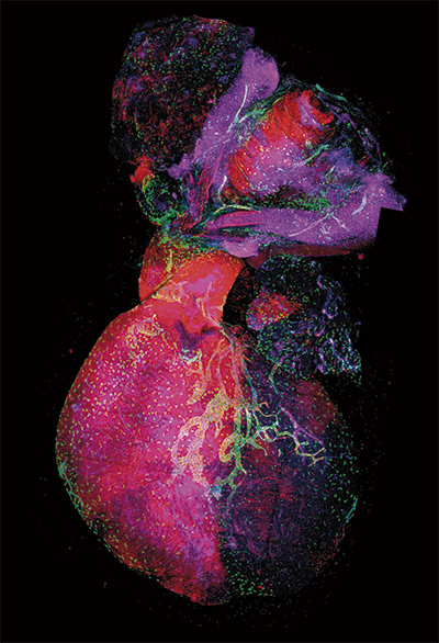
A new way to make arteries
A study carried out at the CNIC not only advanced understanding of the biology of blood vessels, but also pointed the way to the design of new therapeutic strategies to induce vascularization and more effective blood perfusion of injured or ischemic tissues.
The study, led by Rui Benedito and published in Nature, reveals a new cellular and molecular mechanism essential for the development of arteries from blood capillaries, a process called arterialization. Activation of this mechanism could improve the recovery of heart function after transient or long term reduction in heart blood flow, as occurs after a heart attack.
The results also have important implications for the use of drugs to boost angiogenesis—the formation of new blood vessels—in ischemic cardiovascular disease.
The study suggests that pro-angiogenic drugs that stimulate general blood vessel proliferation will suppress arterialization. “One of our future goals will be to identify new ways to suppress proliferation signals exclusively in pre-arterial cells, thereby promoting effective arterialization without negatively interfering with the induction of capillary angiogenesis,” said Dr. Benedito.
From a translational standpoint, the authors conclude that the ability to modulate arterial or venous identity of blood vessels is of great interest for the treatment of coronary artery disease and myocardial infarction. The results obtained could lead to novel therapeutic approaches to induce effective arterialization in ischemic cardiovascular disease.
ELIFE
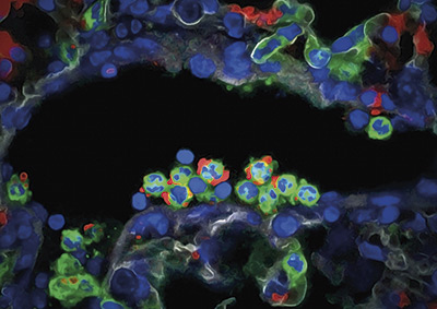
Neutrophils, the hidden controllers of the liver’s internal clock
CNIC scientists have discovered a mechanism underlying the development of steatosis, or fatty liver, one of the main risk factors for liver cancer. The study, published in eLife, for the first time shows that neutrophils act as “circadian messengers” in the liver, controlling its internal clock and lipid metabolism. The study opens a window for potential therapies to treat liver diseases.
The study findings establish that neutrophils are active and crucial circadian regulators of liver metabolism. This could provide a new therapeutic target for the treatment of metabolic diseases such as steatosis, which commonly progresses to certain types of liver cancer.
The study was supported by funding from the Asociación Española Contra el Cáncer.

Science Advances
The role of the CCT protein in the control of immune-synapse formation
A new study published in Science Advances identifies the role of the cytosolic chaperone protein CCT in the reorganization of the cytoskeleton during the formation of an immune synapse.
The research teams led by Francisco Sánchez-Madrid and Noa Martín at the CNIC and the IIS Princesa and by José María Valpuesta at the CNB-CSIC have shown that CCT controls changes in the arrangement of the centrosome and mitochondria by regulating the production of the cytoskeletal components α- and β-tubulin.
This finding opens the path to the design of strategies aimed at blocking CCT for the treatment of autoimmune diseases that involve hyperactivation of lymphoid cells.
NATURE
A new diagnostic and therapeutic target for cardiovascular disease
A study published in Nature showed that the mitochondrial protein ALDH4A1 is an autoantigen involved in atherosclerosis. The CNIC research team concluded that ADH4A1 is a potential marker for the diagnosis and treatment of cardiovascular disease (CVD).
Atherosclerosis develops for many years without causing symptoms, and there is therefore a pressing need for new tools for diagnosis and therapy. Study leader Dr. Almudena Ramiro explained that, “we know that atherosclerosis includes an immunological component and that the innate and adaptive immune systems are both involved in the origin and progression of this disease.” However, little is known about the specific response of B cells in these processes or the repertoire of antibodies these cells produce during atherosclerosis.
Autoantigens are molecules produced by the body that, through a variety of mechanisms, are recognized as foreign and trigger an immune response. “ALDH4A1 is recognized by the protective antibodies produced during atherosclerosis, making it a possible therapeutic target or diagnostic marker for this disease,” commented Ramiro
The study shows that ALDH4A1 accumulates in plaques and that its plasma concentration is elevated in the atherosclerosis-prone mice and in human patients with carotid atherosclerosis, establishing ALDH4A1 as a possible biomarker of the disease.
The team also found that infusion of A12 antibodies into the atherosclerosis-prone mice delayed plaque formation and reduced the circulating levels of free cholesterol and LDL, suggesting that anti-ALDH4A1 antibodies have therapeutic potential in the protection against atherosclerosis.
These results broaden knowledge of the humoral response during atherosclerosis and highlight the potential of ALDH4A1 as a new biomarker and of A12 as a therapeutic agent for this disease.
The study was supported by funding from the “la Caixa” Foundation, the Asociación Española Contra el Cáncer, and the Ministerio de Ciencia, Innovación y Universidades.
Nature Cell Biology
A subgroup of stem cells resists aging and maintains muscle regeneration capacity until geriatric age
Researchers at Pompeu Fabra University, the CNIC, ICREA, and Ciberned have identified a physiological mechanism that maintains the regenerative capacity of muscle stem cells, and surprisingly resists the passage of time far more than expected, until geriatric age.
Skeletal muscle regeneration depends on a muscle stem cell population (satellite cells) in a dormant or quiescent state, a situation that can be triggered by damage or stress to form new muscle fibers and expand in new stem cells.
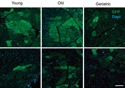
The regenerative functions of these stem cells are known to decline with aging. Dr. Pura Muñoz-Cánoves and colleagues have found in experiments with mice that all muscle stem cells, despite being quiescent, are not equal, and have identified a subgroup that maintains its regenerative capacity over time, declining only at geriatric age.
The researchers have shown that this subgroup of quiescent stem cells has a greater regenerative capacity through the activation of the FoxO signaling pathway (previously associated with longevity), which maintains the expression of a youthful gene program throughout life; however, at geriatric age, FoxO activation in this subgroup of cells is lost, causing their loss of functionality.
The results show that compounds that activate FoxO can have a rejuvenating effect on aged muscle stem cells, opening the way to improving the health of elderly people affected by the loss of muscle mass. The research may also benefit people who have lost muscle mass as a result of neuromuscular diseases or effects associated with cancer or infectious or inflammatory diseases.
The study was supported by funding from the “la Caixa” Foundation, the ERC, and the Ministerio de Ciencia e Innovación y Universidades.
Cell
The unexpected repair function of neutrophils
The CNIC team lead by Dr. Andrés Hidalgo has discovered that neutrophils, the most abundant cells of the innate immune system, have many more functions in the body than previously thought. This finding suggests possible new treatments for many diseases, including cancer.
In a study published in the journal Cell, the research team demonstrate that neutrophils acquire new characteristics when they arrive in a tissue and that these specialized functions help to maintain organ health.
Every day, the marrow inside our bones produces immense quantities of neutrophils. These cells then enter the bloodstream and are distributed to almost all tissues of the body. Neutrophils have a short lifespan, living for less than 24 hours. For this reason, scientists believed that these cells had a very limited capacity to adapt to their environment and adopt new functions.
But the authors of the Cell study found that when neutrophils leave the circulation and migrate into tissues they acquire new, previously unknown properties.
According to Dr. Hidalgo, the most fascinating finding is that neutrophils appear to acquire functions useful to the specific tissues in each organ.
This ability to change cell properties was identified in healthy individuals, which suggests that neutrophils participate in a great variety of normal functions in the body and are not limited to combating infection.
Previous studies had already identified neutrophil heterogeneity in several diseases. Indeed, these neutrophil changes are prognostic markers in cancer and help to regenerate blood cells after bone marrow transplantation.
However, the mechanisms that establish neutrophil hyperplasticity are poorly understood, and the new results are a crucial step towards filling this knowledge gap. Essentially, what we have demonstrated is that neutrophils, despite their sort lifespan, can change their function and that they do this when they enter tissues. The identification of these adaptations allows a better understanding of the roles of different immune cells in disease.
The study was supported by funding from the European Regional Development Fund (ERDF), RTI2018-095497-B-I00, the “la Caixa” Foundation, and Leducq Foundation Transatlantic Network of Excellence (TNE-18CVD04).
Nature Reviews Endocrinology
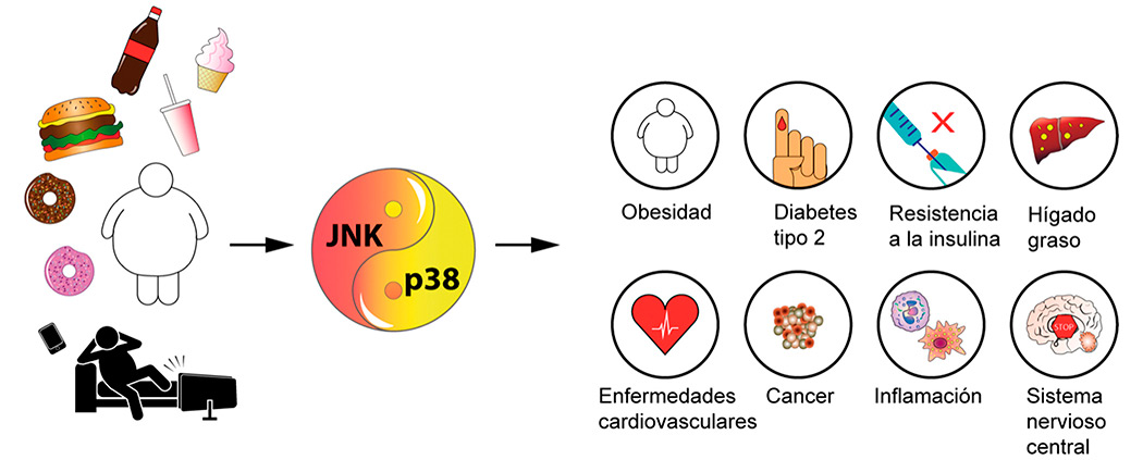
Stress kinases could be the next target in the treatment of obesity-associated diseases
CNIC investigators from the group led by Dr. Guadalupe Sabio reviewed the key role played by stress kinases in adjusting metabolism to changing conditions in an article published in Nature Reviews Endocrinology.
Stress kinases are proteins in the body that are associated with obesity and metabolic alterations such as insulin resistance and diabetes. Understanding their mechanisms and routes of action could be crucial for the design of therapeutic strategies to combat the global epidemic of obesity, which affects millions of people worldwide.
JNK and p38 SAPKs are known to act through diverse signaling pathways and mechanisms that affect metabolic processes such as insulin sensitivity, thermogenesis, and lipolysis. In addition, SAPK activity in immune cells triggers inflammation in several tissues and alters systemic metabolism in obesity. The accumulated evidence also shows that SAPKs have tissue and substrate specific functions and that their activation is involved in disorders related to obesity, steatohepatitis, liver cancer, heart failure, and diabetes mellitus.
The review concludes that the main goal in this field is to identify specific inhibitors for each of the individual p38 and JNK members. These molecules can then be tested for their ability to prevent and treat obesity and associated diseases.

Dr. Sabio’s group is supported by funding from the Fundación Científica de la Asociación Española Contra el Cáncer, a BBVA Beca Leonardo para Investigadores y Creadores Culturales (2017), and the European Foundation for the Study of Diabetes.
Science
A new cellular defense mechanism against viral and bacterial infection
A study published in the journal Science and coordinated by researchers at the CNIC, IDIBAPS, and the University of Barcelona describes a hitherto unknown immune defense mechanism orchestrated by lipid droplets (LDs), cellular organelles able to attract and eliminate invading pathogens.
LDs accumulate fatty nutrients that provide the energy cells need to perform their functions. For example, LDs provide the energy for the heart to beat, the liver to perform its metabolic functions, and muscles to move.
The newly identified mechanism highlights the intimate relationship between metabolism and immunity, and could stimulate the development of new therapeutic strategies at an especially critical time, when antibiotic resistance is presenting an ever increasing threat to public health.
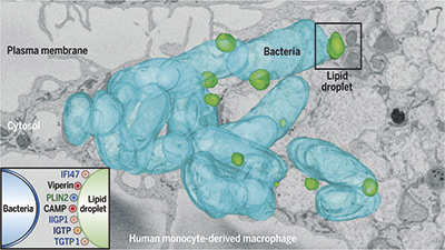
This study represents a paradigm shift because until now LDs were thought to be at the service of viruses or bacteria during infection. “In light of the widespread resistance to current antibiotics, this study has deciphered an important defense mechanism that could be stimulate the development of new therapeutic strategies to stop infections.”
The study was supported by funding from the Human Frontier Science Program Organization.
Angewandte Chemie International Edition
Controlling the speed of enzyme motors brings biomedical applications of nanorobots closer
A study by scientists at the CNIC, Universidad de Girona (UdG), and the Institute for Bioengineering of Catalonia (IBEC) has overcome one of the key hurdles to the use of nanorobots powered by lipases.
The study was coordinated by CNIC investigator Marco Filice and was published in the journal Angewandte Chemie International Edition. The study describes a tool for modulating motors powered by enzymes.
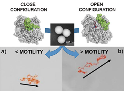
Microorganisms are able to swim through complex environments, respond to their surroundings, and organize themselves autonomously. Inspired by these abilities, over the past 20 years scientists have managed to artificially replicate these tiny swimmers, first at the macro-micro scale and then at the nano scale, finding applications in environmental remediation and biomedicine.
The speed, load-bearing capacity, and ease of surface functionalization of micro and nanomotors has seen recent research advances convert these devices into promising instruments for solving many biomedical problems. However, a key challenge to the wider use of these nanorobots is choosing an appropriate motor to propel them.
The authors conclude that this new tool for modulating enzyme-powered motors broadens their potential biomedical and environmental applications.
EHJ
Metoprolol: an old drug with unique cardioprotective properties
CNIC researchers have demonstrated the unique properties of metoprolol, a treatment able to reduce the long term consequences of a heart attack. Metoprolol, a member of the beta-blocker class of drugs that has been in use for more than 40 years, has been found to have unique cardioprotective properties.
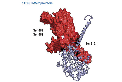
This is the conclusion of a study carried out by scientists at the CNIC, Fundación Jiménez Díaz University Hospital, and CIBERcv. The study, performed in sophisticated experimental mouse models, shows that the cardioprotective effect of metoprolol during a heart attack is not shared by other beta-blockers commonly administered by intravenous injection, such as atenolol and propranolol.
The European Heart Journal study, led by Dr. Borja Ibáñez, “demonstrates that metoprolol has unique cardioprotective properties and heralds a paradigm change in cardiology and the treatment of acute myocardial infarction.”
The authors conclude that metoprolol should be the beta-blocker of choice in clinical practice. “If these results are confirmed in future clinical studies, this would herald a change in the clinical guidelines for this devastating disease, placing metoproplol, but not other beta-blockers, as the drug of choice for patients suffering a heart attack,” said Dr. Ibáñez.
JACC
Vigorous exercise, spongy heart
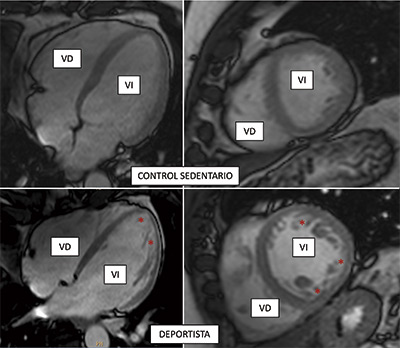
Scientists at the CNIC and CIBERcv have used cardiac magnetic resonance technology to measure exercise-related hypertrabeculation in a general, non-athlete population. The results of the study have important practical implications because “misdiagnosis of noncompaction cardiomyopathy in people who exercise regularly (whether professional athletes or amateurs) can trigger medical recommendations to stop physical exercise unnecessarily,” explained CNIC General Director Valentín Fuster.
The authors conclude that cardiac magnetic resonance criteria for diagnosing noncompaction cardiomyopathy should not be interpreted in isolation. Instead, imaging results should be placed in the context of other clinical parameters, genetic tests, and the level of physical activity. This is important even in a population of non-athletes in order to avoid misdiagnosis of the disease. Misdiagnosis can result in the unnecessary cessation of exercise, with all its associated negative physical and psychological consequences.
The study, published in the Journal of American College of Cardiology (JACC), forms part of the PESA-CNIC-Santander study.
Circulation Research
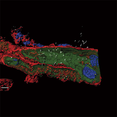
An essential factor for the correct formation of the coronary arteries
CNIC scientists have discovered a protein that participates in the maturation of the blood vessels that supply the heart. Loss of this protein during embryonic development leads to an anomalous remodeling of the coronary arteries. The study, published in Circulation Research, provides the basis for further research into clinical applications to promote vascular regeneration after coronary injury.
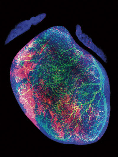
The study, carried out in a rodent model, focused on the embryonic formation of the coronary tree, the blood vessel system that supplies the heart muscle. The research team used a range of transgenic mouse lines and high-resolution microscopy techniques to obtain an unprecedented visualization of the different phases of coronary vascularization.
The researchers used a range of fluorescent genetic indicators, each one targeting a distinct regulatory element of the gene nestin, to isolate different subtypes of endothelial cells—the cells that line blood vessels—before and during the remodeling of the immature vascular network.
By comparing the molecular profiles of each endothelial cell network, the team identified the restricted presence of the transcription factor Sox17 in vessels undergoing arterial remodeling. Specific elimination of Sox17 from the coronary endothelium produced a severe phenotype characterized principally be defective formation of the left coronary artery.
JACC
A new algorithm for personalized cardiovascular risk estimation in healthy people
CNIC scientists have designed an algorithm that provides a personalized estimate of cardiovascular risk in healthy middle-aged individuals based on a range of variables including age, blood pressure, diet, and blood and urines markers. The EN-PESA algorithm is an affordable tool for estimating the severity of subclinical atherosclerosis—characterized by the deposition of fatty substances in the arterial walls—especially in individuals at higher risk. The researchers conclude that EN-PESA will “help to personalize the estimation of cardiovascular risk, leading to tailored treatments and follow-up plans.” The study is published in the Journal of American College of Cardiology.
The success of machine learning algorithms derives from the analysis and systematic processing of huge quantities of data collected from a large number of individuals. One of the pioneering studies in this area is PESA-CNIC-Santander, led by Dr. Valentín Fuster.
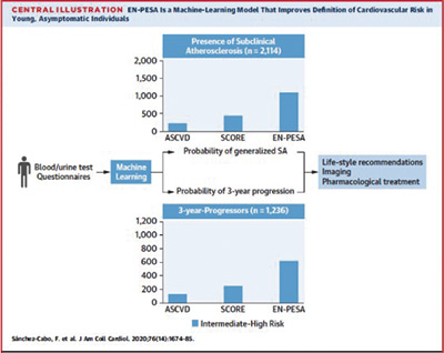
Sifting through this immense quantity of information, the EN-PESA algorithm identified a small set of variables that are easily measured in primary care centers. These variables accurately predict the extent of subclinical atherosclerosis and disease progression in middle-aged individuals who are classed at low or intermediate risk according to established cardiovascular risk scales.
The parameters include age, blood pressure, data collected in routine blood and urine analysis, and answers to dietary questionnaires.
The authors concluded that “this algorithm will improve the clinical management of apparently healthy individuals with a low cardiovascular risk according to established risk scores but who have a generalized extent of subclinical atherosclerosis or a high short-term risk of significant disease progression.”
Nature Metabolism
How immune cells regulate obesity
Two CNIC groups have described how macrophages regulate obesity in a study published in Nature Metabolism that could lead to the design of treatments for obesity and overweight, as well as for related conditions such as fatty liver disease and type 2 diabetes.
The study, led by Drs José Antonio Enríquez and David Sancho, explains how fatty tissue inflammation and obesity emerge from the activation of the mitochondrial metabolism in macrophages in response to oxidative stress due to excess nutrients.
The research analyzed how macrophage metabolic changes regulate the inflammatory process underlying obesity and the metabolic syndrome. The new findings reveal how the detection by macrophages of oxidative danger signals—known as reactive oxygen species—leads to mitochondrial metabolism changes in these immune cells essential for their differentiation to the proinflammatory M1 macrophage type. This oxidative stress is found in morbidly obese patients and seems to be related to the high-fat diet typical in developed economies.
A key finding of the study is that a reduction of this oxidative stress ameliorates some of the harmful parameters associated with obesity.
The results support the potential use of specific Fgr protein inhibitors to treat patients with obesity or metabolic syndrome. The goal would be to reduce the inflammation associated with these conditions, reducing impact of associated diseases fatty liver and type 2 diabetes and contributing to increased life expectancy and quality-of-life.
The study was supported by funding from the Human Frontier Science Program Organization (HFSP RGP0016/2018).
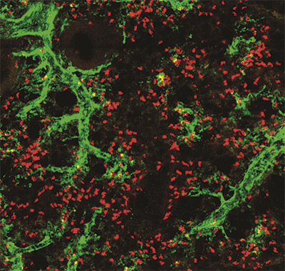
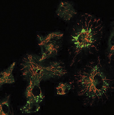
Cell
A cell-cleaning system keeps hearts healthy
Two CNIC groups, led by Drs Andrés Hidalgo and José Antonio Enríquez, have discovered a cell-cleaning system that is key to keeping a healthy heart. This mechanism allows the heart’s contractile cells (the cardiomyocytes) to release damaged components outside the cell into particles called exophers. These exophers are then taken up by a network of immune cells living inside the heart-the macrophages, which are in charge of removing them before they cause inflammation in the heart.
The study, published in Cell, compiles the results of over five years of research, with collaborations with laboratories in Europe, Asia and the US. The insights that this study provides suggests that cardiac dysfunction can emerge in some instances from defects in resident immune cells, rather than from cardiomyocytes. This finding has important implications for the diagnosis and treatment of heart disease.
The finding that cardiomyocytes surrogate the removal of their waste to macrophages has many implications. “The fact that the heart requires a population of macrophages to do their cleaning work, among other tasks, suggests that many heart diseases of unknown origin can be explained by failure of these macrophages,” said Dr. Enríquez.
Another potential implication is that there may be similar processes supporting the fitness of specialized cells in other tissues, including the brain, whose cells share features with cardiomyocytes.
The authors conclude that the identification and elimination of cardiomyocyte-derived mitochondria and other material by resident macrophages establishes a new paradigm of how resident phagocytes contribute to general tissue maintenance.
JACC
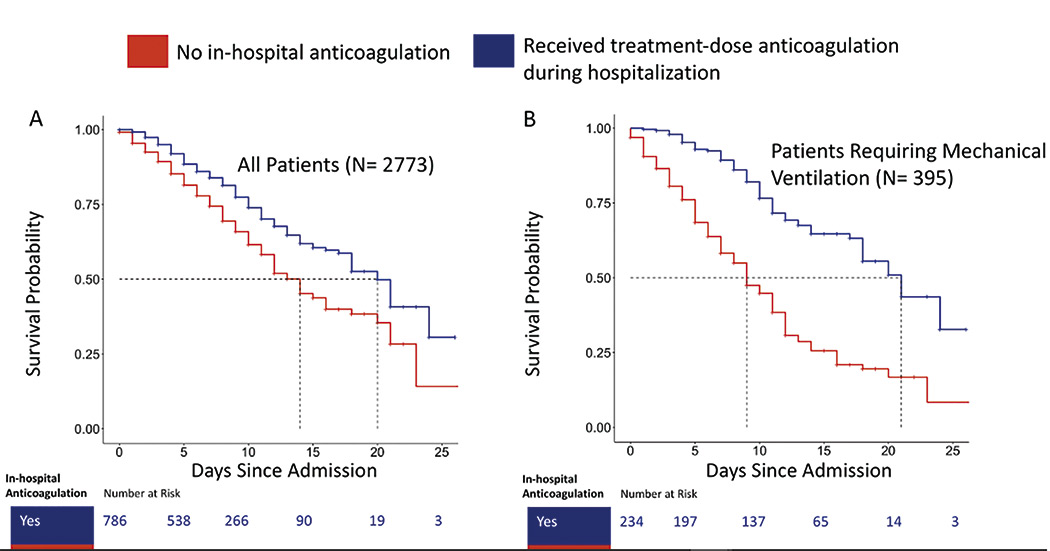
Additional data on the efficacy of blood thinners in COVID-19 patients
Anticoagulation therapy improves survival among hospitalized COVID-19 patients by helping to prevent fatal events associated with coronavirus, such as infarction and stroke. The study, published in the Journal of the American College of Cardiology and led by CNIC Director Dr. Valentín Fuster, shows that hospitalized COVID-19 patients treated with anticoagulants—drugs that prevent blood clotting—had a better survival rate that those who did not.
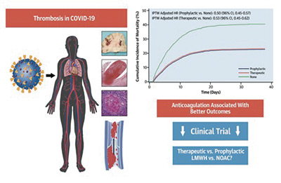
At the start of the pandemic, the same research team demonstrated that anticoagulation therapy was associated with superior survival among hospitalized COVID-19 patients. This subsequent observational study showed that patients on both a “therapeutic” or full dose, and those on a “prophylactic” or lower dose, showed about a 50% higher chance of survival, and roughly a 30% lower chance of intubation, than those not on anticoagulants.
The study examined 6 anticoagulant treatment regimens. Among these, the best outcomes were obtained with therapeutic and prophylactic low-molecular weight heparin and therapeutic apixaban.
Nature
A key mechanism in hypoxia
Researchers at the CNIC and the Instituto de Investigación Sanitaria del Hospital Universitario de La Princesa (IIS Princesa) have made a major advance toward deciphering the mechanism through which the production of reactive oxygen species (ROS) increases in the early phases of hypoxia (acute reduction in tissue oxygen). The finding represents an important advance in the understanding of cell physiology and could be exploited in the future to treat a variety of diseases in which hypoxia plays a role, such as stroke and heart attack. The study was published in Nature.
The study shows that sodium ions act as second messengers that regulate mitochondrial function, specifically the function of the mitochondrial electron transport chain (ETC), by inducing the controlled production of ROS.
This mechanism of ROS production is fundamental to the ability of the pulmonary circulation to respond to hypoxia by redistributing blood flow to regions with less irrigation, a phenomenon known as hypoxic pulmonary vasoconstriction.
Several of the study findings provide important information about cell physiology. First, the study shows that mitochondrial sodium regulates cell membrane fluidity. This was unknown before and could have major implications for the understanding of a multitude of cellular processes.
A second important discovery is that the mitochondrial supercomplexes of the ETC play an important role in this process by adopting structural conformations that are sensitive or insensitive to sodium, thus ensuring that the action of sodium is nontoxic.
Finally, the study shows that inhibition of the mitochondrial Ca2+/Na+ exchanger NCLX is sufficient to block this pathway, preventing hypoxia adaptation. This finding indicates that NCLX could be a future therapeutic target for diseases in which hypoxia plays a role.
The study results reveal that sodium controls OXPHOS function and cell signaling in hypoxia through an unexpected interaction with phospholipids, with major consequences for cell metabolism.
The study was supported by funding from the International Human Frontier Science Program Organization (HFSP RGP0016/2018).
Science Advances
CNIC scientists discover the mechanism of competition between mitochondrial genomes coexisting in the same cell
Research at the CNIC has identified the mechanism of competition between distinct mitochondrial genomes coexisting in the same cell. The study, published today in Science Advances, examines why the simultaneous presence of more than one variant of mitochondrial DNA (mtDNA) in a cell is rejected in most tissues, which select a single mtDNA variant that differs between different tissues.

The results show that cells are able to detect and select between mitochondria with distinct mtDNA variants that make them more or less efficient for different cell types depending on their metabolic needs. This explains how the preferred mtDNA variant can differ between cell types.
The study establishes that the selection of a mtDNA variant depends on the cell type and not the tissue, as was thought previously.
Moreover, the researchers add, the study has identified molecular targets for the development of tools to control mtDNA selection and cell metabolism in order to prevent the accidental generation of heteroplasmy through new medical procedures. These new technologies include the transplantation of healthy mitochondria to prevent mitochondrial diseases, injection of mitochondria into oocytes to increase fertility, and the proposed transfer of mitochondria in cell therapies to treat diverse diseases, including cardiovascular, lung, and neurological diseases.
The study was supported with funding from the International Human Frontier Science Program Organization (HFSP RGP0016/2018).
PNAS
A new mechanism controlling liver cancer development
CNIC researchers have discovered a mechanism controlling the development of a type of liver cancer. This study, published in the Proceedings of the National Academy of Sciences (PNAS), partly funded by the Spanish Association Against Cancer, has identified a protein that, when blocked, dramatically reduces the impact and progression of this type of cancer, called cholangiocarcinoma. This work was possible because CNIC researchers developed an animal model in which alterations in the production of bile acids cause this type of tumor.
In the study, led by Guadalupe Sabio and Alfonso Mora, mice were bred whose livers do not contain the proteins JNK1 and JNK2. The researchers found that these two proteins control the production in the liver of bile acids, which are essential for proper fat digestion and the absorption of fat-soluble vitamins (A, D, E and K). “A lack of JNK1 and JNK2 in the liver leads to changes in the enzymes responsible for metabolizing cholesterol and bile acids,” said Dr. Mora. In the analyzed mice, “we observed excess blood levels of bile acids.”

This model allowed the CNIC team to show that that a key role in this tumor process is played by the protein PPARα, which regulates the metabolism of bile acids and liver lipids. According to Dr. Mora, mice lacking PPARα “have significantly fewer tumors or none at all.”
Although it is still unknown if these data can be extrapolated to human patients, the availability of this first animal model will allow the study of a type of tumor that can still only be diagnosed in its very late stages, when metastases have already occurred.
Earlier studies had shown that JNK blockade prevented the development of steatosis in the liver, leading to a variety of clinical trials with inhibitors of these proteins.
The researchers believe that the new findings are a ‘wake-up call’ for these drugs.
The study was supported by funding from the Asociación Española Contra el Cáncer and a BBVA Beca Leonardo para Investigadores y Creadores Culturales (2017).
Science Advances
The mechanism that regulates mitochondrial energy production
CNIC scientists have identified the molecular mechanism by which mitochondria—the main source of the cell’s energy supply—regulate their function to optimize energy production in order to meet the body’s changing needs. The discovery, published in the journal Science Advances, helps to explain how the body regulates its metabolism.
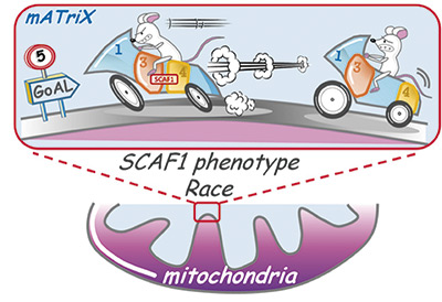
The study, led by CNIC scientists Jesús Vázquez and José Antonio Enríquez, shows that mitochondria adjust the efficiency of the electron transport chain (ETC) to meet the body’s needs by regulating the associations between its component macromolecular structures. The researchers found that the protein SCAF1, discovered by the same team in 2016, plays a key role in this metabolic regulation by optimizing mitochondrial energy efficiency in response to high energy demand.
The research team conclude that SCAF1-mediated physical interaction between CIII and CIV is essential for optimal mitochondrial energy production. SCAF1 is a regulatory factor that allows mitochondria to adapt to the available nutrient sources of sugars, fats, or proteins. This capacity for metabolic adaptation also explains the ability of mitochondria to adapt to stress situations, for example during intense physical exercise.
The study was supported by funding from the International Human Frontier Science Program Organization (HFSP RGP0016/2018), Fundació Marató.TV3, and the “la Caixa” Foundation.
Science Advances
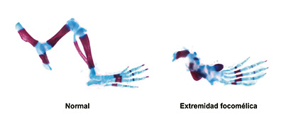
Spanish scientists discover a system essential for limb formation during embryonic development
Researchers at the CNIC have discovered a system that provides cells with information about their position within developing organs. This system, studied in developing limbs, tells cells what anatomical structure they need to form within the organ. The article, published in Science Advances, shows that malfunctioning of this system causes congenital malformations and could in part explain the effect of thalidomide, a drug contraindicated in pregnancy because it induces limb defects.
The CNIC team, working with partners at the National Institutes of Health in the USA, analyzed the molecular basis of limb formation. The scientists discovered how cells obtain information about their position on the proximodistal axis of the limb bud, or primordium (the rudimentary state of a developing organ.)
The study shows that the signal that tells cells where they are is the growth factor FGF. The strength of the signal received by cells depends on how close they are to the FGF-producing cells at the distal tip of the limb bud.
The researchers demonstrated that the molecule in receptor cells that interprets FGF signals is a transcription factor called Meis. This transcription factor is distributed in a linear abundance gradient, so that it is highly abundant in proximal cells (close to the body trunk) and becomes progressively less abundant in more distal positions.
This system, emphasized Torres, “is essential for the correct formation of the limbs.” The mechanisms described in the study advance understanding of the origin of phocomelia, a congenital defect in which the embryonic limbs only form hands and feet, with the rest of the limb failing to develop. In the study, experimental elimination of FGF–Meis signaling resulted in all limb-bud cells receiving the incorrect instruction that they were distal, resulting in phocomelia.
The findings establish a new model for the generation of proximodistal identity in the developing vertebrate limb and provide a molecular mechanism for the interpretation of FGF gradients during axial patterning in vertebrate embryos.
Science
Old or stressed immune cells can damage tissues and accelerate age-associated diseases
Scientists at the Instituto de Investigación del Hospital Universitario 12 de Octubre -i+12- and the CBM, in collaboration with researchers at the CNIC, have demonstrated that an aged immune or stressed immune system can attack the body’s own tissues and accelerate the appearance of several diseases associated with aging.
Published in Science, the results show that when T lymphocytes age or are subject to stress the release a cytokine storm that can affect a number of different organs and tissue, activating a cell aging program known as senescence. The appearance of senescent cells in these tissues predisposes to several conditions, including cardiovascular, neuroinflammatory, metabolic, and muscle diseases.
The most striking finding was that mice with a stressed immune system began to show signs of premature aging and to develop age-related conditions, most notably cardiovascular changes and weight loss. The animals later lost muscle strength and eventually underwent changes in memory and behavior.
In a second part of the study, the researchers sought to identify treatments able to slow the appearance of these symptoms. They used two strategies, one aimed at blocking the cytokine storm and the other at preventing tissue senescence. Both strategies delayed the appearance of disease symptoms.
JACC
Blood thinners may improve survival among hospitalized COVID-19 patients
Anticoagulation therapy improves survival among hospitalized COVID-19 patients by helping to prevent fatal events associated with coronavirus, such as infarction and stroke. The study, led by Dr. Valentín Fuster, Director of the CNIC and the Heart Institute and Mount Sinai Hospital in New York, shows that hospitalized COVID-19 patients treated with anticoagulants—drugs that prevent blood clotting—had a better survival rate that those who did not.
The researchers evaluated the electronic medical records of 4389 patients with a confirmed COVID-19 diagnosis, hospitalized at 5 hospitals between March 1 and April 30, 2020. The team analyzed the survival and mortality rates of patients who received therapeutic or prophylactic doses of anticoagulants (oral antiplatelet drugs, subcutaneous low-molecular weight heparin, or intravenous heparin) compared with the rates for patients who did not receive these treatments.
Overall, 467 (10.6%) of the patients required intubation and mechanical ventilation during their hospitalization. Those on therapeutic blood thinners had 31% fewer intubations than those not on blood thinners, while those on prophylactic blood thinners had 28% fewer. The difference between the two anticoagulant groups was not statistically significant.
Cardiovascular Research
Spanish scientists discover how the flu virus injures the heart
A study by a consortium of Spanish research groups has described the potentially fatal cardiac effects of infection with the influenza virus, especially more virulent forms. The research team showed that the replication of flu virus particles infecting the heart blocks the transmission of cardiac electrical impulses, which can lead to death. The results, published in Cardiovascular Research, highlight the potential value of testing patients with acute cardiac disease for flu infection during seasonal outbreaks of the disease. The study was carried out by scientists at the CNIC, CSIC, CIBERES, CIBERCV and the Hospital Clínico San Carlos together with scientists at the Universities of Michigan and South Florida in the United States.
The investigators assessed the mechanisms of heart injury in mice infected with the flu virus as well as in cardiac muscle cells derived from human pluripotent cells. The results show that the most virulent viral strain persists in the heart for longer and triggers changes, including the modification of ion channels responsible for the transmission of cardiac electrical impulses. These results provide a molecular explanation for the early death of infected mice and suggest that screening for infection in acute cardiac disease patients during seasonal flu outbreaks could enable early diagnosis and treatment.
The authors concluded that “these findings highlight the potential benefit of early detection of the virus in patients with acute heart disease during flu outbreaks.”
Nature Communications
CNIC scientists design an experimental mouse model for investigating the mechanical function of proteins in vivo
The CNIC Molecular Mechanics of the Cardiovascular System group, led by Jorge Alegre Cebollada in partnership with an international scientific team, has generated the first experimental mouse model that allows direct analysis of the mechanical function of proteins in living organisms.
The model, published in Nature Communications, is based on the insertion of the HaloTag-TEV module into titin, one of the proteins responsible for the elasticity of skeletal and cardiac muscle. The HaloTag-TEV module combines three key properties. Dr. Alegre explained: “Thanks to the introduction of this module into the gene, we are able to fluorescently mark the protein, which makes it easy to track the module and see where it has inserted correctly.”
The module also includes a target for specific protein lysis, so that the mechanical function of the target protein can be interrupted in a controlled manner at any desired moment, allowing us to study the effect of this interruption. Finally, the module provides a way to anchor the isolated protein to surfaces, enabling the study of its mechanical properties in isolation.
“All of this helps to establish a bridge between the modulation of protein mechanical properties and observing the consequences of this modulation at a cellular level,” said Dr. Alegre.
The HaloTag-TEV module can be inserted into other proteins with a mechanical function, so that in the future it could be used to study other systems, including those related to diverse muscular and cardiac disorders.
JACC
Spanish scientists identify a biomarker that detects atherosclerosis before the appearance of symptoms
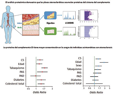
Scientists at the CNIC and the Instituto de Investigación Sanitaria-Fundación Jiménez Díaz (IIS-FJD) in Madrid have demonstrated that a proteins present in early atheroma plaques—accumulations of cholesterol in the walls of arteries—could be used as a biomarker to detect atherosclerosis in the subclinical phase, before the appearance of symptoms.
The study, published in the Journal of American College of Cardiology (JACC), concludes that activation of the complement system is one of the most characteristic molecular changes taking place in the early development of atherosclerotic plaques. The study shows that plasma levels of the complement system protein C5 could be used to identify individuals with subclinical atherosclerosis in a noninvasive way and at minimal cost.
“This early identification of the disease would help to select individuals who would benefit from more costly noninvasive imaging analysis to get a more precise estimate of their cardiovascular risk,” explained Dr. Jesús Vázquez, head of the Cardiovascular Proteomics Laboratory at the CNIC and one of the coordinators of the study.
The study was supported by the Comunidad de Madrid (Complemento II-CM, S2017/BMD-3673) and the “la Caixa” Foundation (HR17-00247).
JACC
Atherosclerosis progresses rapidly in healthy people from the age of 40
Almost half of apparently healthy people between the ages of 40 and 50 could be accumulating fatty plaques—atheromas—in their arteries at a much faster rate than was previously thought. the Journal of American College of Cardiology (JACC) has today published new data from the PESA-CNIC-Santander study demonstrating that atheroma plaques extend rapidly through the arteries of 40% of asymptomatic individuals aged between 40 and 50 years.
Researchers at the CNIC, led by Dr. Valentín Fuster, Director of the CNIC and lead investigator on the PESA-CNIC-Santander study, have also found that the progression of atherosclerosis is directly related to classical cardiovascular risk factors: age, sex, hypertension, cholesterol, smoking, and diabetes.
The researchers conclude that the findings, although they await validation from future events in the PESA cohort, will be of great value for identifying strategies to stall the cardiovascular disease epidemic.
Nature Communications
CNIC scientists discover a new molecular mechanism that regulates the sentinel cells of the immune system
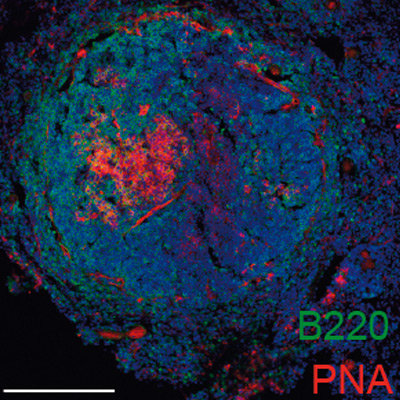
A team at the CNIC, working in partnership with researchers at Mount Sinai Hospital in New York, has discovered a new molecular mechanism mediated by nuclear receptors that determines the identity and expansion of macrophages—one of the cell types that act as immune sentinels in the body. The newly discovered mechanism specifically affects the macrophages resident in the serous cavities, the membrane-surrounded cavities that enclose and protect many organs. The findings, published today in Nature Communications, could have important implications for the treatment of diseases that affect the serous cavities and the organs they contain, including many cancers and myocardial infarction.
There are three serous membranes: the peritoneum, which covers the abdominal cavity; the pleura, surrounding the lungs; and the pericardium, which covers the heart. “One of the main functions of the macrophages residing in these cavities is to maintain homeostasis by removing dead cells,” explained study coordinator Dr. Mercedes Ricote. In addition, recent studies have demonstrated that these macrophages can infiltrate adjacent injured organs, “generating an effective rapid repair response that is independent of the recruitment of macrophage precursors via the blood supply.”
The study demonstrates that the expansion of peritoneal macrophages after birth and their maintenance during adult life are controlled by retinoid X receptor (RXR), a member of the nuclear receptor family.
Using models of ovarian cancer in mice, the study shows that peritoneal macrophages can infiltrate ovarian tumors and act as ‘tumor-associated macrophages’ that “support tumor growth,” explained Dr. Ricote.
The findings demonstrate that loss of RXR function leads to a decrease in the number of macrophages in the peritoneal cavity, resulting in a decreased contribution of these macrophages to growing ovarian tumors, slowing the progression of the disease.
The researchers are especially interested in the possibility of modulating RXR with drugs, including some that are currently used to treat cutaneous lymphomas. “Our research could have implications for the treatment of diseases in which serous cavity macrophages contribute to disease progression, such as cancer, or to the repair of damaged tissue, as in myocardial infarction.”
Journal of Experimental Medicine
An immunological regulatory circuit may play a central role in ocular inflammatory disorders
CNIC scientists have identified an inflammatory regulatory circuit in the eye controlled by a subtype of endothelial cells, the cells that line the interior of blood vessels. The discovery was made by analyzing gene expression in 8,000 cells of the choroid—the vascular layer at the rear of the eye between the retina and the sclera. The results, published today in the Journal of Experimental Medicine, open new perspectives on the study and treatment of retinal vascular disease and inflammatory disorders that affect the choroid.
The study provides valuable information about how the endothelial cells in the choroid regulate the development of inflammatory and vascular diseases in the retina. The authors analyzed the choroids of adult mice using the ‘single cell RNAseq’ technique, which allows analysis of gene expression simultaneously in thousands of individual cells.
Nature Immunology
Neutrophils are equipped with a ‘disarmament’ program that prevents the immune system going ‘out of control’
Scientists at the CNIC have discovered a ‘disarmament’ mechanism that protects our bodies against uncontrolled activity of the immune system. This newly identified immune control system is located in one of the most important cell types of the immune system, the neutrophil. The findings, published in Nature Immunology, could have major implications for the understanding and treatment of conditions such as myocardial infarction, stroke, and acute inflammation.
The study identifies an intrinsic cell mechanism that modifies the protein content of circulating neutrophils, leading to the progressive loss of the toxic contents of the granules and therefore a reduced capacity to form NETs. This program thus undermines the main offensive mechanism of neutrophils. The study shows that this disarmament program is driven by the receptor CXCR2 and regulators of circadian rhythms.
The findings show that neutrophils possess “an innate system that gradually reduces their capacity to mount a toxic attack, so that as they age, neutrophils disarm themselves before they can damage healthy tissues. There are many diseases that cause more or less damage according to the time of day, and this disarmament program helps to explain the origin of these clinical differences,” explained the researchers.
Development Cell
A CNIC-led international study discovers a new origin of lymphatic vessels in the heart
The study, published in Development Cell, has identified and characterized a new vasculogenic niche that contributes to the development of the cardiac lymphatic system. The study shows that the coronary lymphatic vessels have varied origins and functions: the results of the study reveal that the coronary lymphatic vasculature does not have a single origin, but instead forms through the participation of cells from different tissues.
This study opens the way to future research into the mechanism underlying lymphatic vasculogenesis in this new niche and the functional diversity of coronary lymphatics.
This international study, led by Dr. Miguel Torres’s group at the CNIC, examined the origin of the coronary lymphatic system during the formation of the heart in the mouse embryo. The study shows that the heart contains a second population of lymphatic cells that is recruited later during development and is derived not from veins, but from a region called the second heart field.
The second heart field is composed of multipotent cells “able to generate different types of heart cells, including cardiomyocytes (the cells of the cardiac muscle), smooth muscle cells, and the endothelial cells of the arteries and veins.”
One of the more surprising findings was that lymphatic cells generated in the second heart field mix with lymphatic cells with a different, likely venous, origin; the two populations together form the lymphatic vessels in the ventral part of the heart.
This result indicates that the newly discovered cell population not only contributes a large proportion of the cells of the coronary lymphatic system, but also leads a specific and irreplaceable process in the formation of the coronary lymphatic vasculature. “This function reveals, for the first time, the specialization of endothelial subpopulations in the formation of the coronary vasculature and opens the way toward a better understanding of the formation of lymphatic vessels, a process essential not only for embryonic heart development but also for the response of the heart to stress and disease in adulthood.”
Nature Communications
The ‘airbag’ that protects cells against stress
CNIC scientists have identified the molecular mechanisms that allow our cells to adapt to, protect themselves against, and survive mechanical stress. The results, published in Nature Communications, show that our cells produce molecules that act as a type of ‘airbag’ in response to mechanical stress. Without this protective and adaptive system, the heart, an organ subject to continuous mechanical forces, “would be unable to correctly perform its blood-pumping role,” explained lead author Miguel Ángel del Pozo.
Human cells are able to perceive, adapt to and respond to mechanical forces. As >Dr. Del Pozo explained, “these forces can sometimes be excessive, placing cells under a mechanical stress that can rupture the cell membrane and result in the death of affected cells. To avoid this rupture and thus prevent cell death, nature has evolved molecular sensors that ‘switch on’ in response to these forces and initiate adaptive and protective processes. The purpose of this response is to adapt cells to these forces before they cause tissue or organ damage.”
The Nature Communications study identified relatively large folded or wrinkled structures surrounding cells that can unfold or flatten when the cell is stretched, thus giving cells an extra coating that prevents breakage upon excessive stretching. “It can be likened to an accordion, which unfolds as it is stretched, thus preventing it from breaking when pulled,” explained the researchers. These folds thus function as a kind of ‘airbag’, cushioning cells against excessive mechanical stress.
The team also identified molecules that participate in this mechanism, permitting cells to perceive mechanical force and initiate the biochemical changes needed to adapt to mechanical stress.
Through partnership with CNIC scientist Jorge Alegre-Cebollada, the team identified molecules with opposing functions; “one of the molecules (FBP17) protects the cell against mechanical stress, whereas another (ABL) makes the cell more sensitive to these forces,” explained the scientists.
Both molecules, working in an ordered fashion, coordinate changes in the cell envelope that protect the cell and the cell skeleton, giving it the structure and solidity needed to resist mechanical stress.
The authors also managed to alter the amount or activity of these molecules in human cells; inhibiting the action of ABL increased protection against mechanical stress, whereas inhibition of FBP17 made cells more sensitive.
The findings are important because knowledge about how cells are protected against mechanical stress “will give us a better understanding of the molecular basis of diseases such as some forms of muscular dystrophy, cardiomyopathies, and lung or vascular diseases characterized by sensitivity to physical activity. The findings will also shed light on the mechanisms of injury to organs with a high level of mechanical activity, such as the heart, lungs, muscles, and blood vessels.” The authors concluded that “this study opens the way to future therapies in patients with these conditions.”
Europace
Personalized medicine for atrial fibrillation
Patients with atrial fibrillation, the most frequent cardiac arrhythmia, are closer to accessing personalized medicine. This is the claim of a new study led by Dr. David Filgueiras, of the CNIC, the Hospital Clínico San Carlos de Madrid, and the Centro de Investigación en Red de Enfermedades Cardiovasculares (CIBERcv). The study results show that it is possible to monitor and predict individual progression of atrial fibrillation from cardiac electrical signals obtained from implantable devices (pacemakers or defibrillators).
The study, published in the latest edition of Europace, involved the participation of 51 Spanish hospitals and the Fundación Interhospitalaria para la Investigación Cardiovascular. The results show that cardiac electrical signals from patients fitted with pacemakers or implantable cardioverter–defibrillators can be used to monitor and predict the progression of the arrhythmia in a personalized and specific manner. This is achieved with standard data transmission technology installed in implantable devices such as pacemakers. This technology can be used to monitor cardiac electrical activity during episodes of atrial fibrillation, thus establishing disease status and the rate of progression.
According to Dr. Filgueiras, “this technology opens up enormous possibilities in personalized medicine for atrial fibrillation patients because it allows us to determine the progression rate of the arrhythmia in each individual and to optimize the timing of medical intervention with current treatment options.”
In addition, Dr. Julián Villacastín, director of the Instituto Cardiovascular del Hospital Clínico San Carlos and an author on the study, this new approach to atrial fibrillation diagnosis will allow physicians “to monitor the influence of different interventions on disease progression.”
EMBO Molecular Medicine
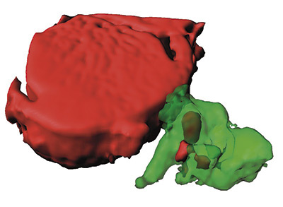
Inhibiting a protease could improve the treatment of inflammatory bowel disease
Scientists at the CNIC and the CSIC have identified the protease MT1-MMP as a possible future target for drugs to treat inflammatory bowel disease (IBD). The study, led by Dr. Alicia G. Arroyo and published in EMBO Molecular Medicine, shows that inhibition of this protease could improve the treatment of IBD.
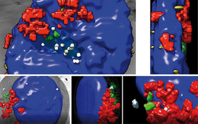
During colitis, the intestinal blood vessels duplicate through mechanisms that are poorly understood. In the new study, Dr. Arroyo’s team used microscopy techniques and 3D image analysis to characterize these duplication events in a mouse model of colitis. These tools enabled the scientists to demonstrate that MT1-MMP expressed on endothelial cells lining the blood vessels impedes their duplication in the inflamed gut, reducing the severity of colitis.
This finding has potential clinical implications. “The study shows that patients with mild IBD have higher than normal circulating levels of TSP1, which could be a useful biomarker of the disease,” commented Arroyo.
In addition, the team managed to reduce vessel duplication in mice with colitis by administering either an antibody that inhibits MT1-MMP protease action or a TSP1 peptide that blocks TSP1-αvβ3 binding. This result establishes the MT1-MMP-TSP1-αvβ3 integrin pathway as a new therapeutic target, particularly for less severe forms of IBD.
The authors concluded that “the study presents a new opportunity to develop personalized treatments not only for patients with IBD, but also for patients with other diseases that progress through vessel duplication, like cancer.”