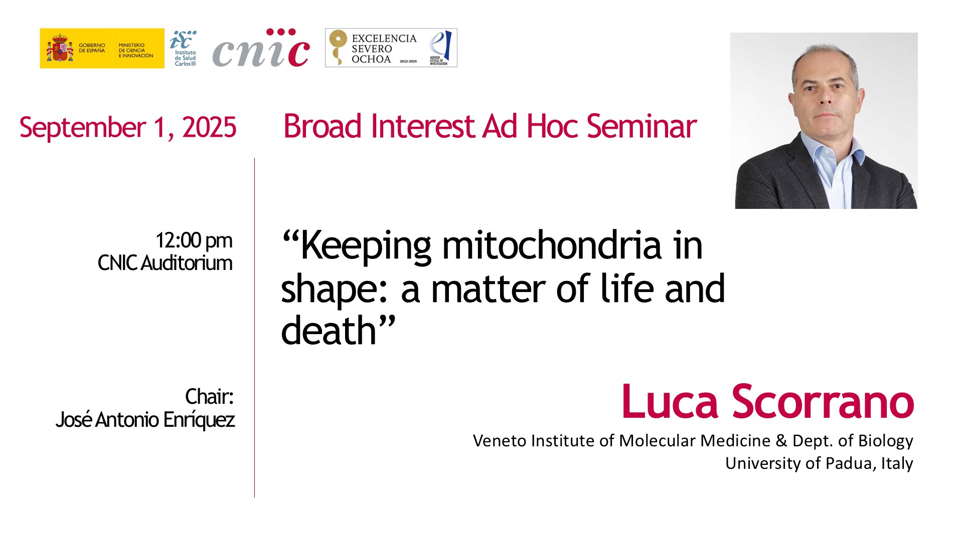
Luca Scorrano: "My fascination with mitochondria has been like a first love”
Luca Scorrano, M.D., Ph.D., is a Professor of Biochemistry at Padova University Medical School (Italy) whose work explores how mitochondrial structure dictates cellular function. His laboratory pioneered the discovery of cristae remodeling and mitochondrial dynamics, identified the MFN2/Ermit tether between ER and mitochondria, and revealed how mitochondrial shape controls processes from apoptosis and stem cell differentiation to heart development, progesterone synthesis, and defense against infection. Scorrano’s research has uncovered mechanisms behind optic atrophy, angiogenesis, adipocyte browning, and mitochondrial disorders, and his findings have led to potential targeted therapies. He has authored 231 papers, is a Clarivate Highly Cited Researcher (2021, 2022, 2024), EMBO member (2012), and Academia Europea member (2019).
- You trained as an MD. Have you ever practiced clinically?
I completed all the clinical requirements, and I’m still registered as an MD in Italy, so technically I could practice. But it’s better for patients that I don’t.
- So, you didn’t enjoy practicing medicine?
Not exactly. That’s why I love mitochondria. I found basic science far more intellectually stimulating. At the University of Padua, where I did my MD, there’s a long tradition in mitochondrial research, so joining a mitochondrial lab felt natural.
- How did you start working with mitochondria?
I initially started in a molecular oncology lab, but I didn’t find it very interesting. While still a medical student, I began visiting Paolo Bernardi’s lab, who later became my PhD mentor. After completing my MD and clinical rotations, I joined the PhD program under his supervision in the Department of Biomedical Sciences. That’s when I started doing hardcore basic science—bioenergetics, studying how mitochondria convert energy from food and regulate ion fluxes across the inner mitochondrial membrane.
- What fascinated you most about mitochondria?
During my PhD, I became fascinated by the role of mitochondria in programmed cell death—a field that was very prominent in the late 1990s. It was astonishing to see that this tiny cellular organelle, essential for converting food into usable energy, is also central to apoptosis, or cellular suicide. In other words, mitochondria are a double-edged sword: indispensable for life, yet equally essential for death.
In 1996, Xiaodong Wang made a remarkable discovery: a component of the same machinery used for ATP production—oxidative phosphorylation—also initiates apoptosis. This was a striking example of how nature repurposes the same system for multiple functions. Inspired by this, I went to Harvard Medical School to join Stan Korsmeyer’s lab, where I studied how mitochondria change shape during these processes. Korsmeyer, a founding father of apoptosis research, had discovered most of the genetic regulators of cell death.
- How has your understanding of mitochondria evolved over the years?
Over the years, I’ve seen mitochondria evolve in our understanding—from “ATP factories” to central regulators of inflammation, cellular recycling, stem cell maintenance, metabolism, and many other cellular processes. The cardiovascular system is no exception: mitochondria are critical not only for producing the ATP needed for heart contraction, but also for vascular regulation, smooth muscle contraction, stem cell differentiation into cardiomyocytes, and angiogenesis. In my view, changes in mitochondrial shape are just as important as their bioenergetic functions.
My fascination with mitochondria has been like a “first love”—initially sparked by their role in energy metabolism, then deepened by their central role in regulating fundamental cellular processes. Over time, my research has expanded to include communication between mitochondria and other organelles. But, as with a first love, you never forget it—and I haven’t.
- How do mitochondria evolve and maintain these functions?
This is a fascinating concept. Evolutionary, mitochondria are descendants of archaeobacteria that invaded primordial cells. It’s a bit more complicated than that, but essentially, this parasitic relationship became mutually beneficial. Mitochondria could harness metabolites from the host cell, and their ATP production was far more efficient than glycolysis alone.
However, there was a challenge: these bacteria had their own replication machinery, which involved both fusion and division. Over evolutionary time, the host cell gained control by losing the bacterial genes responsible for division and instead using proteins—already employed to regulate other membrane systems—to control mitochondrial shape and behavior. This makes perfect evolutionary sense: the invading organelle provided a benefit but also posed a threat, which the host mitigated by integrating mitochondrial regulation into existing cellular signaling pathways.
This control is precise: the cell can direct mitochondria to specific locations, coordinate their division with the cell cycle, and manage asymmetric partitioning during division. But mitochondria are not passive—they can “take revenge.” If the cell damages their membranes, mitochondria release proteins that trigger cell death. If both inner and outer membranes are compromised, they release mitochondrial DNA, which the cell interprets as a viral infection, triggering inflammation.
Thus, control comes with a price. The organelle contains “venoms” that can harm the cell or organism if mismanaged. It’s a truly stimulating example of symbiosis: mitochondria are domesticated yet retain the capacity to defend themselves. During intracellular infections, mitochondria dynamically change shape to control invaders, serving as the first line of defense, while pathogens can manipulate them to escape at the right moment.
Studying mitochondrial morphology reveals the heart of this evolutionary battle between host and parasite. Perhaps the host has “won,” but only partially—evolution is never final. There is no ultimate victory, only ongoing adaptation.
- You mentioned that mitochondrial shape and dynamics are involved not only in the cardiovascular system but also in cancer, infection, and pregnancy.
Yes, it’s a complex topic. For example, during pregnancy, one critical moment occurs around the third month, when the site of hormone production needs to shift. Remarkably, all cholesterol-derived hormones—testosterone, progesterone, and estrogen—pass through intermediates produced in mitochondria. This means mitochondria are essential for sexual reproduction: without the right mitochondrial enzymes or the proper cholesterol supply at the correct time, sex hormone production would fail.
In mammals and birds, mitochondrial regulation of hormone synthesis is crucial for species conservation. At the level of the placenta’s syncytiotrophoblast, changes in mitochondrial shape ensure cholesterol delivery is precise, supporting progesterone production and maintaining pregnancy. Human pregnancy is intrinsically inefficient: only about 30% of unprotected intercourse at peak fertility results in a child, and most miscarriages occur around this critical third-month switch—exactly when mitochondrial shape changes are most important.
Problems with mitochondrial dynamics have also been linked to preeclampsia and eclampsia, which are major causes of maternal morbidity and mortality. While the exact mechanisms are not fully understood, these examples highlight how central mitochondria are across multiple biological processes.
Unfortunately, research into mitochondrial roles in pregnancy has lagged behind cardiovascular research, partly because it has been historically considered a “women’s issue.” Cardiovascular diseases, which primarily affect men in later life, have been studied more extensively. Nevertheless, we now know that mitochondrial shape and dynamics are critical for cardioprotection, determining the extent of damage during ischemia-reperfusion, remodeling the heart in cardiomyopathy, and supporting angiogenesis.
- How does this knowledge help in understanding mitochondrial diseases?
Mitochondrial diseases are among the most prevalent genetic disorders. Although they have diverse genetic causes, they often affect the same cellular pathways—similar to how different types of cancer impact common mechanisms yet have unique features. We are only now beginning to understand in depth how mitochondria function and what goes wrong with these disorders.
Research is challenging because there are relatively few patients, making clinical trials difficult, and funding is limited, so most work falls on academic laboratories. Despite these obstacles, we have a responsibility to improve patients’ lives. Currently, treatment is mostly supportive, which is frustrating, but I believe that in the next 10–15 years, breakthroughs will provide therapies that substantially improve quality of life.
Much like cancer, each mitochondrial disorder may require tailored therapies. To achieve this, we must first understand the fundamental principles of mitochondrial function and dysfunction. Our goal is not only to extend life but also to ensure patients can live with dignity. I remain closely connected with many mitochondrial disease patients and the associations that support them, which continually reinforces the urgency and importance of this work.


