News search

E.g., 17/04/2024
E.g., 17/04/2024
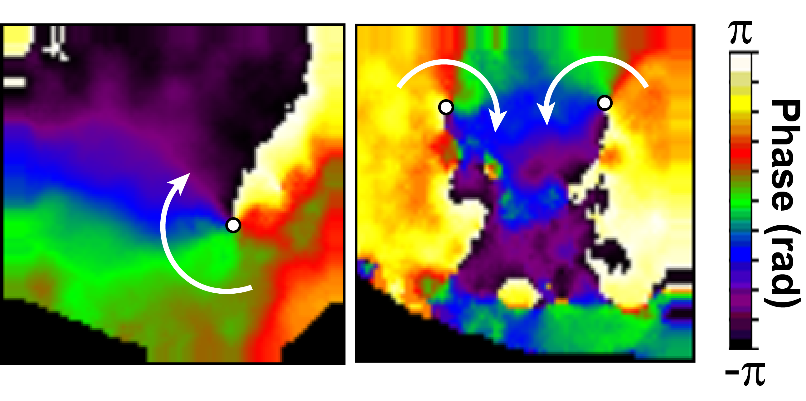
Imágenes de mapeado óptico de dos corazones distintos de ratones que desarrollaron fibrilación ventricular.
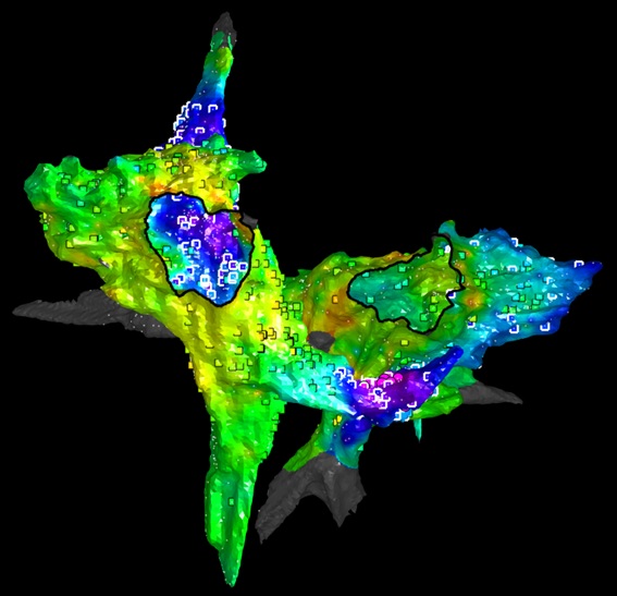
Atrial electroanatomic map of a pig with persistent AFib. The regions colored magenta, purple, and dark blue have high mean FM values. These regions maintain the AFib, and their ablation with a catheter stops the arrhythmia. The squares represent the sites of rotational activity, which are sensitive but not specific to the regions that maintain the fibrillation.
Research
12 Sep 2019

The new study was conducted by investigators at the CNIC, the Hospital Clínico San Carlos in Madrid, and the CiberCV research network and is featured on the cover of the latest edition of the journal Circulation Research