News search
 About the CNIC 18 Mar 2025 The CNIC has been allocated €405,625 for its involvement in GRACE, a project focused on developing innovative strategies to improve the detection and management of cardiovascular diseases. |
 Research 3 Oct 2024 The method, validated in a multicenter study led by the Hospital Clínico San Carlos and the CNIC, enables preoperative planning based on a cardiac magnetic resonance imaging strategy that avoids the biases intrinsic to conventional image analysis |
 Research 3 Mar 2023 The results, published in eClinicalMedicine, have direct implications for clinical practice by providing a list of reference values for a multitude of cardiac parameters used in daily practice |
 Research 22 Jul 2022 Genetic screening combined with the detection of fibrosis identifies patients at risk of malignant arrhythmias or developing heart failure with severe complications |
 Research 28 Sep 2021 This innovative technology may provide an efficient approach in clinical practice after manual or automatic segmentation of myocardial borders in a small number of conventional 2D slices and automatic scar detection |
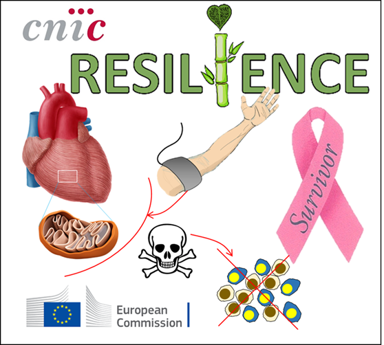 About the CNIC 29 Jun 2021 Over one million European cancer patients suffer from side effects to chemotherapy |
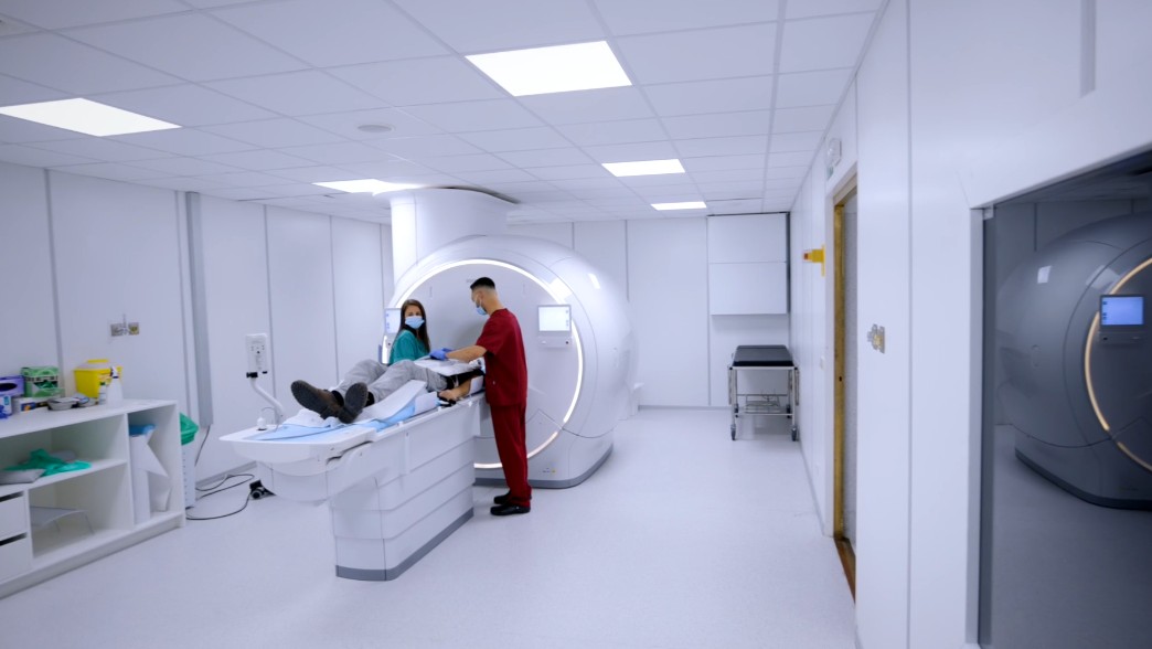
 Research 22 Apr 2021 Ultrafast cardiac magnetic resonance allows precise assessment of heart anatomy and function while reducing healthcare costs and increasing patient comfort |
 Research 7 Apr 2020 A CNIC study published in JACC demonstrates that atheroma plaques extend rapidly in the arteries of asymptomatic individuals aged between 40 and 50 years participating in the PESA-CNIC-Santander study. |


