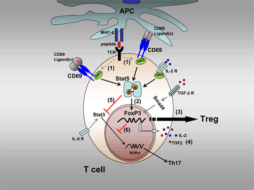
Figure 1: CD69 receptors are expressed on the membrane of T cells following activation (1). The cytoplasmic tail of CD69 associates with Jak3 and Stat 5 proteins, triggering phosphorylation of Stat5 and its translocation to the nucleus (2) where it can activate the transcription factor FoxP3, stimulating the differentiation of regulatory T cells (3). CD69 engagement can also induce expression of IL-2 and TGF-?. These cytokines may act in an autocrine manner to induce the differentiation of regulatory T cells (4). CD69 can inhibit the Th17 differentiation pathway through at least two mechanisms: CD69-activated Stat5 directly inhibits the translocation of Stat3 to the nucleus (5) and indirectly, via FoxP3 activation, antagonizes Stat3-mediated ROR?t activation (6). APC, antigen presenting cell; TCR, T cell receptor; Treg, regulatory T cell; P, phosphorylation.

Figure 2: CD69 acts as a brake on the progression and severity of autoimmune myocarditis and the development of dilated cardiomyopathy (DCM). Our study paves the way to investigations into whether defects in CD69 expression or function influence the development of DCM in humans. These findings increase our knowledge of the development of myocarditis, providing a cellular and molecular basis for the development of novel therapies.
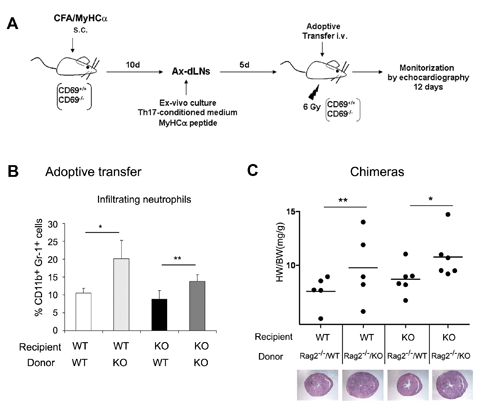
Figure 3: Adoptively transferred CD69-/- Th17 cells can induce severe myocarditis in WT mice. (A) WT and CD69-/- Th17 cells were produced by sensitizing mice to MyHC-? peptide followed by isolation from axillary-draining lymph nodes (AX-dLNs) and in vitro derivation. The Th17 cells were then injected into either WT or CD69-/- recipient mice. (B) Analysis of inflammation in recipient hearts. Bars represent the proportion of infiltrating neutrophils (CD11b+ and Gr-1+) in the myocardium 12 days after Th17 cell transfer. (C) CD69 WT and KO mice were lethally irradiated and reconstituted with a mix of bone marrow cells from RAG2-/- plus CD69-/- or RAG2-/- plus CD69+/+ mice. Heart weight/body weight (HW/BW) ratios of individual chimeric mice after the induction of EAM are shown as dots; horizontal bars represent means. Representative myocardial cross sections are shown below the chart. Data correspond to the arithmetic mean and SD (n=6), and p values are indicated (one-way ANOVA and Bonferroni multiple comparisons test).


