"Going abroad, at least for a while, is highly recommended for everyone, and especially for scientist"


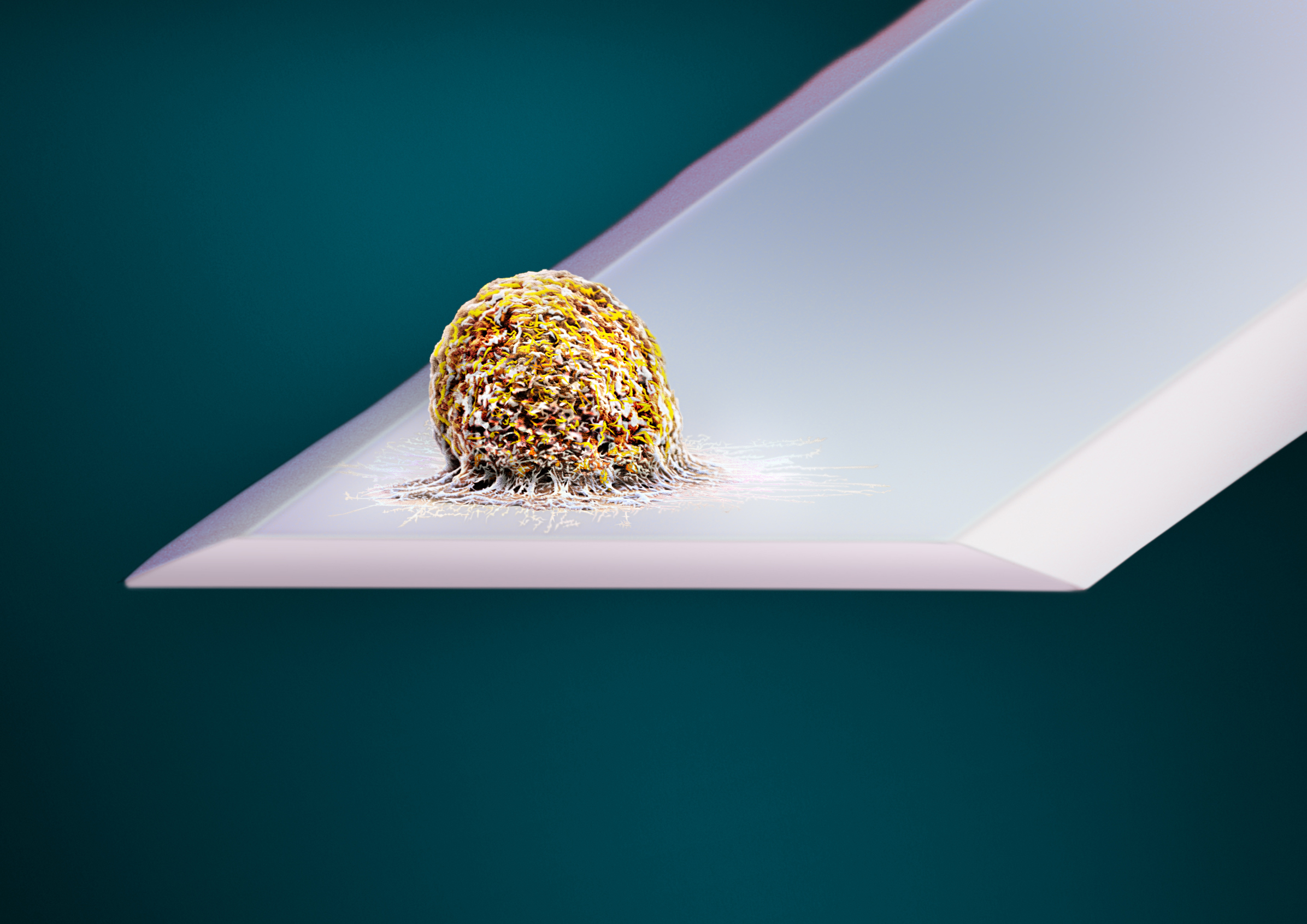
Dr. David Martínez Martín (ETH Zürich, Suiza) has created a technique that allows us to measure in real time the mass changes that cells experience throughout their lifetime
Dr. David Martínez Martín holds a degree in Physics from the University of Valladolid with an Extraordinary End-of-Degree Award, and a PhD in Physics from the Autonomous University of Madrid, obtaining the Prize from the Royal Academy of Doctors of Spain. He has also done some training in the USA, Germany and Switzerland - where he ended up moving to in 2012 to work at the Federal Polytechnic University of Zurich (ETH Zürich), where he continues to develop his research career. With the aim of discovering how cells regulate their mass and size, Dr. Martínez Martín has created a technique that allows us to measure in real time the mass changes that cells experience throughout their lifetime (Nature). This technology makes it possible to measure the mass of individual cells or cell aggregates under culture conditions for days, with a resolution of milliseconds and a mass sensitivity of several picograms (0.1% of the mass of mammalian cells). Using his method, he has observed that the mammalian cells experience subtle mass fluctuations in a matter of seconds, or that the evolution of their mass is different when they are infected by a virus. He is currently preparing his transfer to Australia, where he has recently accepted an offer from the University of Sydney to start up his laboratory there.
- What is your line of research?
I work in biophysics and biomedical engineering. I am very interested in discovering the mechanisms that regulate cell growth. Although, they are considered the most elementary living units, the cells are actually complex autonomous systems. From a thermodynamic point of view, cells are open systems, that is, they are able to exchange energy and mass with their environment. Therefore, any model that tries to explain cellular functioning must include the exchange of mass and energy.
The mass and size of the cells is not random, but rather, it is intimately related to their biological function and physiology. For example, adipocytes in the human body are much larger than fibroblasts, and these in turn are larger than the beta cells of the pancreas. These facts suggest that the cells have mechanisms to regulate their mass and size. These mechanisms are essential in the formation of highly complex organisms such as humans, with a sophisticated architecture of tissues and organs.
To this day, it is known that the origin of many diseases (cancer, hypertrophies, diabetes ...) is related to problems in the regulation of cell mass, so discovering the functioning of these mechanisms is a priority. Nonetheless, although characterizing mass changes of systems much larger or smaller than cells is done routinely, we did not have technologies that would allow this type of measurements at the cellular level with the necessary resolution. That’s why I decided to work on the development of new instrumentation that will allow us to change this situation.
This technology will open new ways to research cellular physiology in greater depth and will allow new diagnostic techniques to be established.
- How does this instrument work?
It is based on a fundamental property of matter, which is inertia. A microscopic arm captures the cell or cells to be studied, and keeps them in culture conditions. With the help of a modulable intensity laser, a very slight oscillatory movement of the arm is induced in the atomic scale, which allows a characteristic frequency to be located that depends on the mass of the cells. A second laser picks up the movement of the microscopic arm and sends it to an electronic system that analyzes it, extracting the information of the cell mass with very high precision.
The instrument is non-invasive and allows us to follow in real time (10 milliseconds of resolution) the evolution of the mass of the cells with a sensitivity of several picograms (approximately 0.1% of the mass of a human average cell). In addition, an inverted optical microscope is incorporated in the device and is fully compatible with transmission optical microscopy, fluorescence, confocal microscopy, etc. This allows simultaneous contrast of cell mass information with morphology and cell state.
- What about the cellular environment?
To guarantee the cellular environment, we have also designed and built an environmental control system that guarantees the pH, osmolarity, temperature, and prevents contamination. This way the measurements can be made in a cultivated environment for days.
- What information have you obtained so far?
A fascinating observation that we have made is that the mass of mammalian cells fluctuates slightly over time. We have detected fluctuations in mass at different time scales, but the fastest ones occur in just a few seconds. It is possible that these fluctuations are a fingerprint of the regulation system of cell mass regulation, although we still do not know its specific origin. The experiments we have conducted so far indicate that they are related to the metabolism and water exchange of cells with their environment.
We have also done research to see if the regulation of cell mass changes when they are infected by a virus. Specifically, we have worked with the Vaccinia virus, and we have discovered that the cells infected with this virus keep their mass close to a constant value, which allows us to easily distinguish between infected and uninfected cells.
Taking into account these results, I am convinced that this technology will open up new ways to investigate the cellular physiology more in depth and will allow new diagnostic techniques to be established. For example, it could be used to develop new antibiotic sensitivity tests. Currently these type of tests take between 24 and 48 hours, but with our technology you could probably get the results in less than 30 minutes. An advance that could save many lives.
- Would it be possible to understand how a healthy cell becomes cancerous?
One of the indications of the formation of a tumor, for example, in the mammary epithelium, is pleomorphism. That is, the manifestation of cells in that tissue with very different sizes. Therefore, I think that we could extract information of great clinical relevance by observing what happens to the mass of the cells during this type of processes.
Currently you are in Switzerland, but you will be moving to Australia. Is it the case of the Spanish scientist in search of an opportunity?
Science is a global activity; there are no borders. When I finished my PhD, I was lucky enough to be able to choose where I wanted to continue my career. I got a scholarship from the international excellence program EMBO (European Molecular Biology Organization) and I moved to Switzerland with the challenge of designing an instrument that would allow us to track the evolution of the mass of cells with great precision.
The project was very risky, a lot of funding was needed and the instrument had to be built from scratch. A project of this kind cannot be executed anywhere, but Switzerland had a very attractive environment. So I decided to go there and work with great scientists like Professor Daniel Müller and Professor Christoph Gerber (Kavli Prize in 2016).
I have been in Switzerland for more than 6 years and I am convinced that going abroad, at least for a while, is highly recommended for everyone, and especially for scientists. It allows you to open up to other cultures, to get to know other ways of working, to master another language. It helps eliminate many prejudices and significantly promotes personal development.
Currently these type of tests take between 24 and 48 hours, but with our technology you could probably get the results in less than 30 minutes. An advance that could save many lives.
- Do you have any plans for collaborating with centers in Spain?
Collaboration with other specialists and other centers is essential. Spain has very good scientists and research centers, and I am currently in contact with several groups to study possible collaborations. In particular, I think that very interesting collaborations could be established with the CNIC.
- How did you become interested in science?
Since I was a child I wanted to be an inventor. I loved the games in which I could build engines, electric circuits, water pumps, etc. In High School, I discovered that I loved mathematics and physics, and that they gave me the power to explain and predict many phenomena. That’s why I decided to study Physics at the University.
- This is your first visit to the CNIC. What’s your impression?
I knew that the CNIC is a center of reference, and having the opportunity to know first-hand the excellent research carried out here has been very rewarding. The CNIC is a great example of how to connect basic research with clinical practice.
- Dr. David Martínez Martín (ETH Zürich, Switzerland) held the seminar "Tracking a cell's mass in real time: a new indicator of cell physiology" at the CNIC, invited by Dr. Jorge Alegre.


