Alzheimer's & Dementia: A new imaging technique locates microthrombi that worsen Alzheimer’s disease, opening the way to personalized anticoagulant therapy


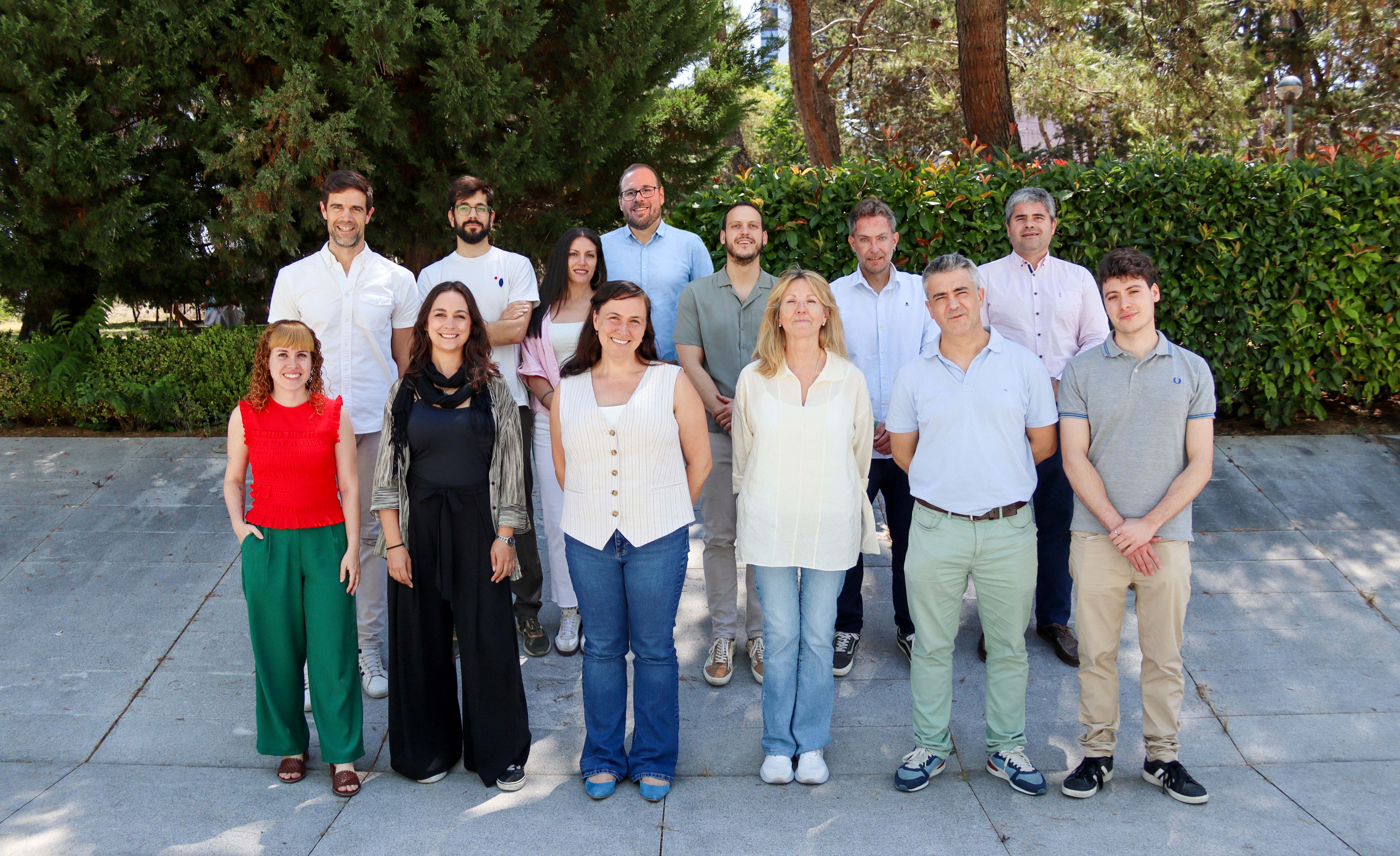
This tool paves the way for identifying patients who would be suitable candidates for treatment with anticoagulant therapy already available at the clinic
More than half of patients with Alzheimer’s disease develop cerebral microthrombi—microscopic blood clots that form in the smallest blood vessels in the brain. This accumulation of microthrombi worsens disease progression. Now, a team of researchers from the Spanish National Center for Cardiovascular Research (CNIC), the Spanish National Research Council (CSIC), and the Fundación Jiménez Díaz Health Research Institute (IIS-FJD) has succeeded for the first time in using noninvasive neuroimaging techniques to detect the accumulation of microthrombi in the brains of living mice modeling Alzheimer’s disease.
The new tool, described in a study published in Alzheimer’s & Dementia, one of the leading international journals in dementia research, opens the way to identifying patients who would benefit from treatment with available anticoagulant therapies, offering a new therapeutic strategy for a disease that still has no cure. Working with partners from institutions including the Gregorio Marañón Health Research Institute, Complutense University of Madrid, and the Center for Energy, Environmental and Technological Research (CIEMAT), the team was able to noninvasively detect a procoagulant state in the Alzheimer’s mouse model.
The team, led by Dr Marta Cortés Canteli, group leader at the Cajal Neuroscience Center (CNC-CSIC) and a collaborating scientist at the CNIC, used positron emission tomography (PET), a medical imaging technique widely used in clinical practice. PET imaging uses radioactive probes that bind specifically to target molecules. In this study, the team used probes that bind fibrin and platelets—key components of microthrombi—to assess accumulation in the brains of the Alzheimer’s model mice: the greater the abundance of microthrombi, the stronger the signal detected by the scanner.
Alzheimer’s disease affects an estimated 55 million people worldwide. It is the leading cause of dementia and a highly complex disorder in which disruption of the brain vasculature plays a central role. More than half of Alzheimer’s patients are estimated to have microthrombi in their brains as a result of an underlying procoagulant state. These microthrombi impair cerebral blood flow and promote disease progression. Although anticoagulant treatments exist that can slow this process, microthrombi have until now remained undetectable until postmortem examination of brain tissue, preventing timely identification of patients who might benefit from such therapies.
Click chemistry
One of the study’s main innovations is its design of PET probes using click chemistry, a groundbreaking approach whose developers were awarded the 2022 Nobel Prize in Chemistry. “This strategy improves image quality and reduces radiation exposure by using a two-step imaging approach: first locating the biological target and then administering the radioactive tracer,” explains Dr Marta Casquero Veiga, a researcher at the IIS-FJD.
Using this strategy, the researchers detected increased probe signal in the brains of Alzheimer’s model mice, supporting a diagnostic approach with strong potential for clinical application. In addition to the findings in mice, the study describes for the first time the presence of platelet deposits in human Alzheimer's disease brain tissue samples, obtained from the Fundación CIEN Brain Tissue Bank. “This finding not only sheds light on the composition of microthrombi and the nature of the procoagulant state in Alzheimer’s disease but also opens the door to new diagnostic and therapeutic targets,” says Dr. Cortés Canteli.
This study aligns with the current multifactorial view of Alzheimer’s disease, which seeks to identify and target the biological mechanisms driving the disease before symptoms appear. “The focus is gradually shifting from clinical manifestations toward the underlying pathological processes, opening new opportunities for early diagnosis and personalized medicine,” says Dr Carlos Cerón. Overall, these results reinforce the potential of noninvasive diagnostic strategies to identify and stratify Alzheimer’s patients based on their biological characteristics, facilitating personalized treatments tailored to each individual.
The study received financial support from the EU Joint Programme – Neurodegenerative Disease Research (grant JPND2020-568-025); the Carlos III Health Institute (ISCIII; grants AC20/00091 and AC20/00077, both within the JPND framework); the BrightFocus Foundation (grant A2023012F); a Juan de la Cierva (JDC) postdoctoral fellowship (JDC2022-048922-I) funded by the Ministry of Science, Innovation, and Universities (MICIU), the State Research Agency (AEI), and the EU through the NextGenerationEU/PRTR programme; the DIAMOND network (grant RED2022-134299-T), funded by the MICIU; the Madrid Nanomedicine in Molecular Imaging Network (Red Madrileña de Nanomedicina en Imagen Molecular, RENIM-CM), funded by the Community of Madrid (grant S2022/BMD-7403); and grants PID2024-157521OB-I00 and CNS2023-144316 to Dr. Cortes-Canteli, funded by the MICIU, the AEI, the ERDF, and the NextGenerationEU/PRTR programme, among other funding.
The project used equipment from the Singular Scientific and Technical Infrastructure (Infraestructura Científica y Técnica Singular) distributed biomedical imaging network (ReDIB), specifically through the TRIMA@CNIC node at the CNIC. This infrastructure is funded by MICIU grant ICT2021-006950 and the NextGenerationEU/PRTR programme.
- Ceron C, Casquero-Veiga M, Lamanna-Rama N, et al. Unveiling the procoagulant state in Alzheimer's disease: A novel PET imaging strategy. Alzheimer's Dement. 2026;22:e71652. https://doi.org/10.1002/alz.71652