CNIC and Philips develop a technique that enables assessment of heart anatomy by MRI in only 15 seconds
The development of this 3D T1 mapping technique, called "SACORA", comes as part of the ongoing collaboration between CNIC and Philips
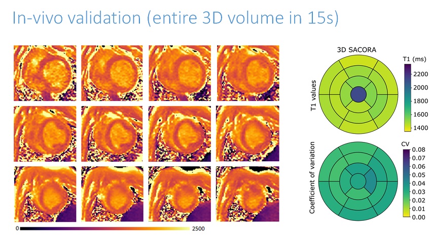
The Spanish National Centre for Cardiovascular Research (CNIC) and Philips, with support of the European project the 4D-Heart (Marie Sklodowska-Curie training programme N722427) coordinated by the CNIC, have developed a technique that makes it possible to obtain information about the composition of cardiac tissue (MRI mapping) in a single apnea, i.e., with the subject holding their breath for 15 seconds.
The new technique to assess the composition of cardiac tissue, called SACORA (SAturation recovery COmpresses SENSE Rapid Acquisition), has been validated in an animal model. The results were published in MAGMA, a specialist journal for magnetic resonance imaging, and allow us "to obtain quantitative data about the composition of cardiac tissue of the whole heart in an acquisition time that is radically shorter than that required for standard T1 mapping", explains Dr Borja Ibáñez, Director of the CNIC Clinical Research Department, cardiologist at the Fundación Jiménez Díaz and member of CIBERCV, the cardiovascular disease biomedical research centre.
Magnetic resonance imaging allows the anatomy (shape) and function (contractions) of the heart to be seen with great precision. Recently, additional magnetic resonance imaging techniques have appeared that allow us to study not only the heart's form and function, but also its composition. "These mapping sequences are the equivalent of a histological study, but are in vivo, and non-invasive", indicates Dr Ibáñez.
It has been shown that the composition of cardiac tissue plays an important role in the propensity to develop future heart problems. The 'T1 mapping' technique allows us to see the composition of the cardiac muscle and, therefore, its use is becoming routine in the study of heart diseases. These techniques provide what are known as T1 mapping that measure an intrinsic property of the tissue, called relaxation time, when exposed to the magnetic field.
The 'T1 mapping' technique allows us to see the composition of the cardiac muscle and, therefore, its use is becoming routine in the study of heart diseases
The presence of areas of microscopic fibrosis, which are not visible with classic methods, can be studied using these T1 mapping techniques. Such diffuse fibrosis is associated with the development of arrhythmias, heart failure and other malignant diseases. Until now, it has been necessary to sweep the heart, taking multiple stills in order to include the whole ventricle and complete T1 mapping of the whole heart, "like slices of bread", explains Ibáñez. With this new method, it is no longer necessary to go "slice by slice" to obtain information about the whole heart, as the whole volume (3D) is acquired in one go.
The development of this 3D T1 mapping technique, called "SACORA", comes as part of the ongoing collaboration between CNIC and Philips. Using SACORA, in apnea (holding air in the lungs in a similar way to when swimming underwater) it is possible to obtain information about the whole of the heart. Dr Sánchez-González, technical leader of the Clinical Science organization in Philips Iberia, points out that otherwise, if ten sections (slices) were needed, it would be necessary to do ten apneas, the study would take a lot longer and consequently the patient would become more tired during the course of the MRI study.
According to Dr Sánchez-González, the last-named author of the paper, SACORA is the "combination of intelligent selection of the acquisition parameters based on the compressed sensing (CS) method of MRI acquisition that gives precise, independently reproducible T1 values independently of the patient's heart rate".
The combination of both advances, in addition to others they are currently working on, may result in full tissue mapping in a greatly reduced period of time, which would revolutionise heart MRI diagnosis
Dr Borja Ibáñez, co-director of the study with Dr Sánchez-González, moreover indicates that, unlike other options where information is obtained from a single heartbeat, SACORA acquires data from 15 beats, and the quality of the image is even better than classic 3D acquisitions where it is necessary to compensate for respiratory movement.
The same research group has developed and patented another revolutionary MRI technique to acquire anatomy and function in a single apnea, called "VF-3D-ESSOS”. The combination of both advances, in addition to others they are currently working on, may result in full tissue mapping in a greatly reduced period of time, which would revolutionise heart MRI diagnosis.


