Developmental Cell: CNIC team reveals how the heart is organized from the earliest stages of embryonic development
CNIC researchers have discovered that the heart is formed from two independent cell types that act in synchrony from the onset of gastrulation. This finding could help to better understand the origin of certain congenital heart defects and open up new opportunities in regenerative medicine and tissue bioengineering.
A study published today in the journal Developmental Cell uncovers new insights into how the heart forms during the earliest stages of embryonic development. The research, led by scientists at the Centro Nacional de Investigaciones Cardiovasculares (CNIC), shows that the heart originates from two distinct cell populations that form independently—but in a coordinated manner—from the very beginning of development: specifically, from the onset of gastrulation, the process by which the embryo begins to organize its basic cellular layers.
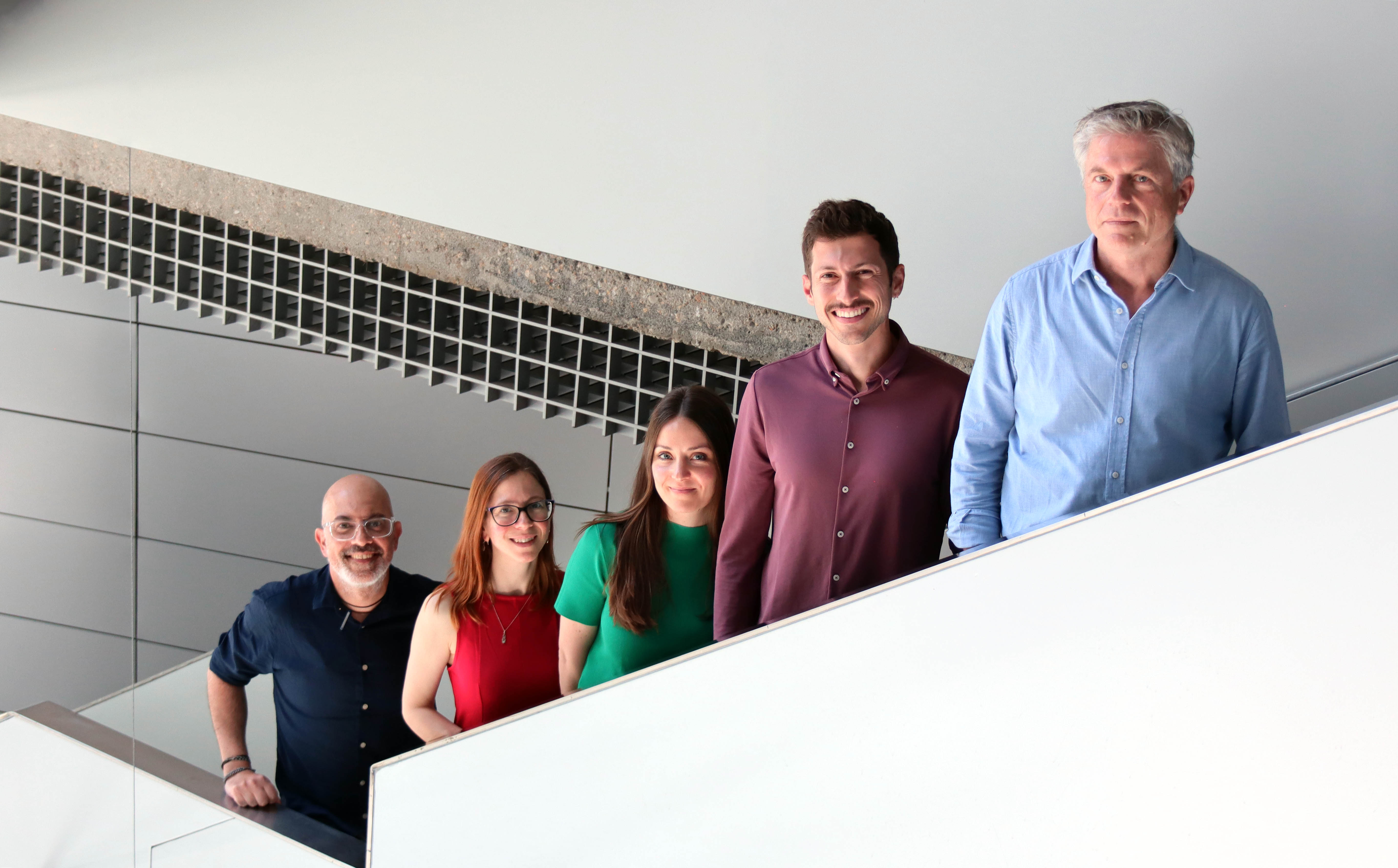
“This discovery is highly significant,” says CNIC scientist Miguel Torres, head of the Genetic Control of Organ Development and Regeneration Group and senior author of the study together with Miquel Sendra. “It helps us understand how the heart is structured in its earliest phases, which may allow us to identify the roots of certain congenital heart defects. It also opens new doors for regenerative medicine and tissue bioengineering.”
Until now, it was thought that cardiomyocytes—the muscle cells of the heart—and endocardial endothelial cells—which line the heart’s inner surfaces—originated from a single precursor population.
However, the new study, based on in vitro mouse embryo culture, advanced microscopy, and cell tracing techniques, reveals that these two cell types originate from separate regions within the mesoderm, one of the three germ layers of the early embryo.
Although they develop separately, the two cell populations enter the embryo simultaneously and migrate in a coordinated fashion toward the region where the primitive heart tube begins to form. According to the researchers, this synchronized behavior points to the presence of highly precise organizational mechanisms at a stage when the embryo still lacks most visible structures.
The study also shows that, despite being fated to form the heart, these cells retain the potential to contribute to the development of other organs—highlighting their versatility and importance in early embryogenesis.
Both lead authors, Miguel Torres and Miquel Sendra, emphasize that this advance was made possible through fundamental research aimed at generating new knowledge without immediate clinical application.
The study was funded by the "la Caixa" Foundation (ID 100010434); the Company of Biologists; the Spanish State Research Agency; the European Commission's H2020 program REANIMA, and CARDIOBOOST-CM of the Community of Madrid.
It also received support from the ERDF ‘A way to build Europe’ (ReDIB ICTS TRIMA@CNIC infrastructure, MCIN).


