Josep Munuera: “Understanding evolution helps us understand today’s diseases”


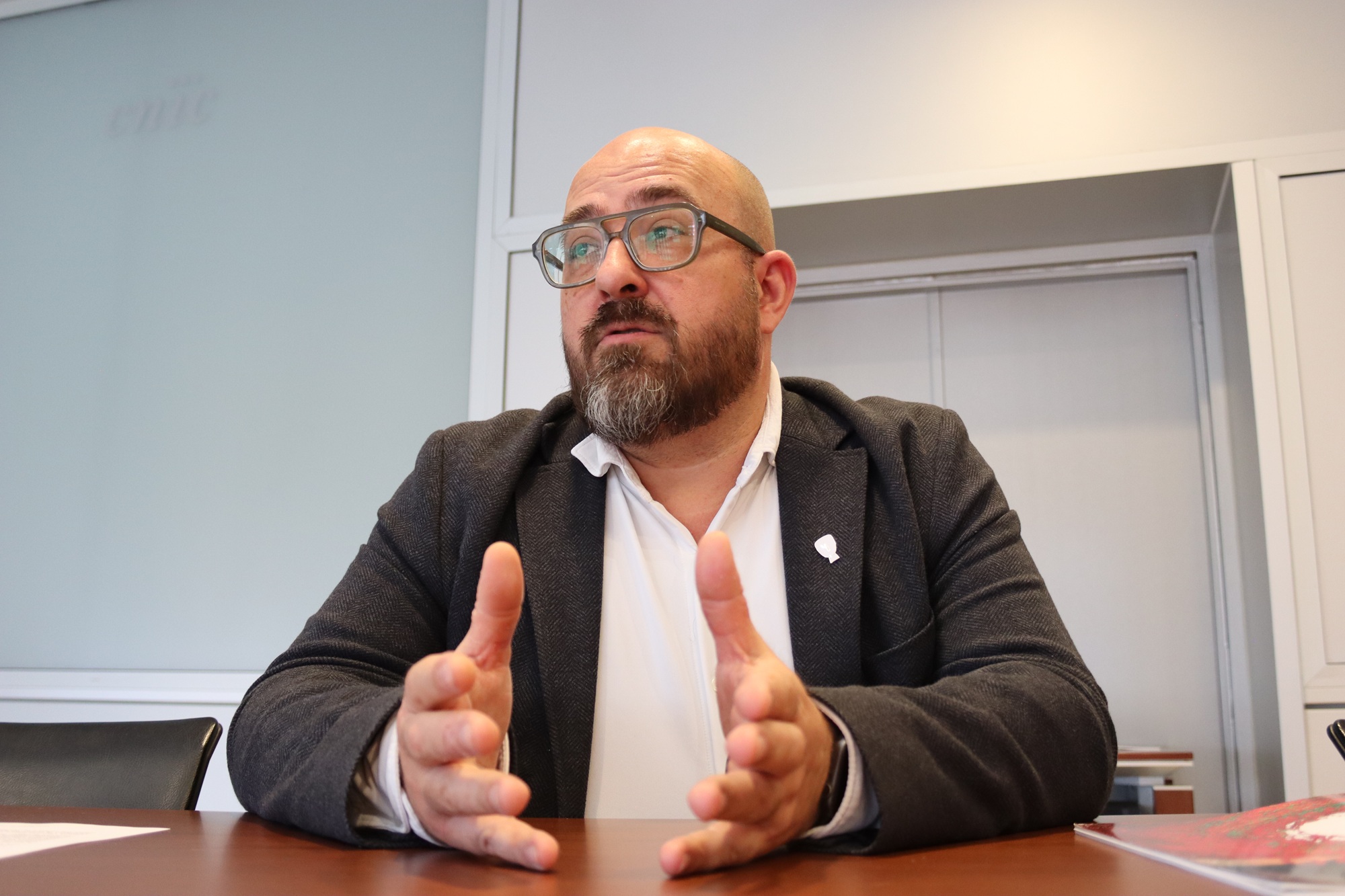
Josep Munuera, Head of the Radiology Department at Hospital de Sant Pau, is a radiologist specialized in neuroradiology—specifically cerebral vascular imaging—and principal investigator in one of the PESA Brain projects. He is also the principal investigator of a Plan Nacional project that uses advanced MRI to quantify cerebral vascular parameters.
- What is the goal of this research?
Essentially, the aim is to extract quantitative data from cerebral blood vessels using flow sequences, magnetic susceptibility, and vascular anatomy, in order to measure vascular characteristics that until now could only be assessed visually—or were simply impossible for the human eye to analyze.
Quantification allows us to obtain numerical data on blood vessels: number, shape, size, diameter, number of branches, flow, velocity, tortuosity, and so on. For example, tortuosity [the degree to which blood vessels deviate from a straight path] is associated with certain cerebral vascular diseases and also with normal brain aging. Until now, all of this was evaluated visually, with limited precision. The trend is to extract these parameters mathematically, so that we can generate comparable metrics and better understand what is happening across different diseases.
- Until recently, neurology and cardiology were considered divergent fields.
And yet, once you understand how a vascular system behaves, it is relatively easy to understand the relationship between different systems. We need to stop thinking of the brain as an isolated system. Blood vessels do not “end” at each anatomical region—the vascular system is a single, continuous network. That is why it should not surprise us that having arterial or venous disorders in the limbs may be associated with an earlier onset of neurodegenerative disease. We need more integrated perspectives.
- Has this new paradigm been fully adopted, or does it still need consolidation?
I think it will take a few more generations for this truly transversal vision to be fully established. It has begun in the basic sciences and in research, and is being adopted more slowly by the medical specialties. In hospitals, traditional structures still weigh heavily—departments, professional silos. The key to breaking down these barriers will probably be functional units—multidisciplinary teams working together.
At the CNIC, more and more groups are moving in this direction. More generally, we perhaps still need to convince some clinicians of the relationship between vascular disease and dementia.
- Can you give an example?
I’ll tell you an anecdote. In 2005, I was working at Vall d’Hebron Hospital performing MRI scans on patients with acute cerebral infarction. Next to me was a cardiac imaging radiologist. I wanted to quickly classify the type of stroke; he said, “It would be useful to know whether it’s of cardioembolic origin.” So we added a “black blood” sequence to obtain a two-chamber image of the heart and see whether there were thrombi in the atrium or atrial appendages. It was very early-stage work, but it already represented a natural integration that allowed us to reach a complete diagnosis in under 15 minutes.
That kind of approach—together with the combined use of imaging data, other omics, and computation—has encouraged a convergence of specialties that previously worked separately or in parallel.
- Or perhaps they are actually the same disease.
We need to explore the origin of diseases from all perspectives. If you treat one aspect without treating the other, you are not really treating the patient. We have to look at the whole person.
- You have that perspective because you work with images and don’t have a clinical bias.
It’s not about bias, but with experience you develop what I would call an ‘agnostic’ approach. In my training, we were taught to look at the image first, almost de novo, without additional information. Only afterwards did we add the clinical layer, to make sense of the finding and correlate it with the patient’s situation. That way of working has stayed with me: looking at the image without preconceptions allows you to generate new hypotheses.
- And this has coincided with the boom in imaging techniques.
Yes, and in that respect I consider myself privileged. I’ve lived through the explosion of advanced imaging, including MRI, CT, PET, and ultrasound. Over the past decade, MRI has become a key tool for understanding anatomical, physiological, and pathological processes—and even aspects of human evolution. And now, with the rise of computation and artificial intelligence, the analytical scope of advanced imaging techniques is expanding even further.
- It’s impossible not to talk about AI.
For me, the key is being able to compute data—ever larger volumes of it—much more quickly. Processing information used to take months; now that time is dramatically reduced, allowing us to devote more time to interpreting relationships between results.
In imaging fields (radiology, pathology, and others), computer vision also allows us to extract information that humans simply cannot interpret—for example, the tortuosity of microvessels measuring 1–3 mm. This was unthinkable twenty years ago.
A few weeks ago, new studies from the BRAIN project were published.
I think BRAIN represented three fundamental things. First, something basic but at the same time revolutionary: collaboration. Building a shared project to collect and share data. That changed the paradigm in many centers.
Second, it coincided with the emergence of advanced MRI, and this allowed us to understand the brain as a fully connected system—the connectome.
And third, it broke down barriers between fields: anatomy, physiology, psychiatry, and technology. That model has since been replicated in different ways, for example in PESA Brain. PESA Brain allows us to study the evolution of a healthy population: normal anatomical variations, their distribution, and how they relate to neurodegenerative changes and cardiovascular risk factors.
- Let’s return to AI. How do you think it will change research?
I see three major areas. First, operations: coding, billing, data transfer, and database cleaning. This might sound like a minor issue, but it will free up an enormous amount of professionals’ time.
Second, data extraction and model generation. We are facing a tsunami of data that we are currently unable to exploit. AI facilitates data analysis, the identification of relationships, and the grouping of profiles. It also allows us to extract information from examinations performed for other purposes. For example, a CT scan done to study lung metastases also contains information about a patient’s metabolic state, coronary arteries, muscle mass, and fat distribution. And in turn, we can relate metabolic status to cancer progression.
Finally, new AI models: synthetic data that will allow us to expand knowledge and databases where data are insufficient. Another emerging field will be hypothesis-driven AI—not only analyzing data, but generating new hypotheses. I’m very curious to see where this takes us.
- And what does evolution have to do with all this?
As a radiologist, my field of knowledge is anatomy—but in digital form. And you cannot understand anatomy without understanding human evolution. The evolution of the skull or of blood vessels can explain why we develop certain diseases.
For example, the size and shape of deep cerebral veins in neonates are related to hemorrhage risk; variants of the cerebral arterial circle are associated with the size of certain infarcts; and the size of venous foramina in the skull influences intracranial pressure and, secondarily, tau protein accumulation, by altering venous and glymphatic drainage. The size of these foramina is directly related to human bipedalism. Understanding evolution helps us understand today’s diseases.
- So understanding our ancestors helps us understand modern disease?
Exactly. The evolution of hominids can explain certain types of disease.