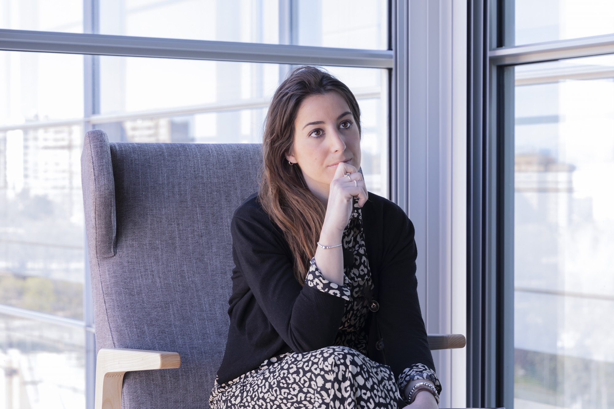
Roser Vento-Tormo: "Working in academic science is a privilege, as it allows us to develop our creative side"
Vento-Tormo completed her PhD with Esteban Ballestar in Barcelona, where she studied the influence of cytokines on innate immune cell differentiation. She later did her post-doc with Sarah Teichmann at the Wellcome Sanger Institute as an EMBO and HFSP Fellow. She was finalist in the 2023 edition of the Michelson Philanthropies & Science Prize for Immunology for her essay “Decoding foreign antigen tolerance: Cell atlases of human tolerogenic milieus guide transformative immunotherapies”. Her research focusses on how cell-cell communication and the tissue micro-environment regulate cell identity and function in the context of immunity and development.
She also designed CellPhoneDB, a novel repository of ligand-receptors and their interactions, and applied CellPhoneDB to study cellular connections from single-cell transcriptomic data.
- Your team is part of the Human Cell Atlas project, which has recently published new results.
Our team is part of the Human Cell Atlas (HCA) international consortium, which aims to create an atlas of all the cell types in the human body at single-cell resolution, including all stages of human development/life cycle in 3 dimensions to obtain a complete view of the human body during health and illness. Recently, one of the initiatives participating in the HCA, called the Human BioMolecular Atlas Programme (HuBMAP) has generated new results. Some of these results were published in three articles in Nature. This research is an excellent resource for researchers like myself who study human biology and diseases, and an essential contribution for the HCA. Understanding the human body at single-cell resolution level will allow us to develop better diagnoses and treatments.
In other words, the goal of HCA is to draw up a cell map of all the cells located in all our organs, first in healthy ones, to later compare them to diseased ones. It’s like having a Google Maps (a map of healthy cells) that also lets us detect deviations from the route (a map of diseased cells).
Technology plays a very important role in this work because, as we progress further, we increase the resolution of results, and in that way obtain more information about the cell and the tissue. So, these cell atlases are developing in parallel with technologies like genomics, bioinformatics, big data, etc. The use of computers is essential because, in the end, it isn’t only about generating data, but about learning to understand them. You have a map, and you have to understand how to read it.
- One of the studies recently published in Nature follows your group’s line of research in the study of placenta cells.
That’s right, in the Greenbaum study, the authors focus on the development of placenta and how the mother’s immune system helps in this process. The work complements a study that my team published some months ago in the same review [Arutyunyan et al. (2023) Nature. doi: 10.1038/s41586-023-05869-0], which is a continuation of my post-doc studies characterising placenta at single-cell resolution [Vento-Tormo et al. (2018) Nature doi: 10.1038/s41586-018-0698-6]. Both papers published this year (Greenbaum et al. and Arutyunyan et al.) help us better understand the vascular reorganisation in the uterus that is necessary to sustain the development of placenta and the embryo. Uterine reorganisation is essential for a pregnancy to continue, and therefore is very important to understand diseases that affect pregnancy, such as preeclampsia.
Our group generates cell atlases of different parts of the human body, with special focus on mucosa-associated lymphoid tissue, to study how they are formed and their abnormalities in different diseases
- What information have the recently published atlases contributed?
The articles published as part of the HuBMAP initiative in Nature describe the cell maps of three organs: the placenta, intestine and kidney. This allows us to obtain views of the different cell types and their organisation in tissues, as well as help us understand the functioning of healthy tissues and those damaged by a disease.
To generate cell atlases, we use a technology called “single-cell resolution transcriptomics”. The transcriptome gives us exclusive information about the cell since, despite almost all the cells in the body of an individual having the same genome, only some genes are active in each of them. The set of genes that are active in a cell is known as the cellular transcriptome. That’s why single-cell resolution transcriptomics is such a powerful tool as it allows us to make inferences about cell identity and function. Because it requires an initial step of tissue digestion, the technology’s greatest limitation is loss of information about the spatial distribution of cells.
To overcome this limitation, we combine single-cell resolution transcriptomics with another technology called spatial transcriptomics. Spatial transcriptomics lets us measure the transcriptome directly on the tissue and therefore obtain the spatial coordinates that each cell occupies in the tissue. In some ways, spatial transcriptomics is as if you did a high resolution histology, contributing exact information about the cells and which genes they express.
An idea doesn’t come from nothing. If you read, you pay attention and think differently, out of the box
Our group generates cell atlases of different parts of the human body, with special focus on mucosa-associated lymphoid tissue to study how they are formed and their abnormalities in different diseases. In our recently published article (Arutyunyan et al. Nature 2023) we analyse the transcriptome of human uterus samples during the first trimester of pregnancy. These samples also contain placenta, a transitory organ formed by the embryo during its development, which plays an essential role in the nutrition, protection and development of the embryo. To do so, the placenta, which surrounds the embryo, is in direct contact with the uterus. Our work allowed us to study communication between the uterus (maternal) and the placenta (foetal) after implantation in humans.
What happens in the period of development after implantation is fascinating. The epithelial cells of the placenta, called trophoblasts, which are of foetal origin, migrate towards the maternal uterine tissue and invade the uterine arteries. This allows remodelling and broadening of the uterine arteries to increase the amount of maternal blood that reaches the placenta, facilitating the exchange of nutrients between the embryo and the mother. This is a unique condition, where foetal and maternal cells share a space to perform a common function: the development of the foetus.
The transformation of the arteries during the first trimester of pregnancy is essential, because abnormalities in this process are related with common problems in pregnancy like preeclampsia or intrauterine growth restriction. What’s more, the migration of trophoblasts has characteristics that are specific to humans, which cannot be reproduced in mice. This may be due to the length of a human pregnancy (9 months) compared to that of mice and, therefore, the need for a much higher amount of nutrients, which are supplied via blood flow.
Our study is really interesting because, for the first time, we define the mechanisms by which trophoblasts migrate to the uterus and maternal arteries. And they key to this is communication between the foetal (trophoblasts) and maternal (uterine) cells.
- How do cells communicate?
Tissues and organs form organised communities. So that each cell does not act individually, coherence and structure are necessary, and for that to happen, they need to speak to each other. There are different spaces in the tissue that specialise in different functions and determine communication hubs of cells. This allows the control of more complex functions, for instance, how much an organ should grow and when the growth should stop.
There are many forms of communication, and many have not yet been studied. One of the most common forms of cell communication, and what we study, is through ligand-receptor interaction. In this case, the cell that sends the signal, secretes or expresses a molecule called a ligand on its surface. This ligand can interact with another molecule called a receptor, which is expressed on the cell surface of another receptor cell of the signal. As these two molecules interact, a signal is activated in the receptor cell, which ultimately activates a specific expression of genes that may have an impact on cell function.
- So, a defect in development means that communication has failed?
It could be due to many reasons, but it is often the case that one of the cells is not talking correctly to the next cell along and so communication is not established, and the cell doesn’t know what to do. Communication guides all of the processes, from a cell that migrates to a place until it proliferates. If there is a defect in this communication, a cell may proliferate more than it should, for instance, and produce something it should not. Or conversely, it doesn’t proliferate, and something doesn’t happen, or goes to the wrong place.
- This approach, performed on healthy cells, serves to study disease?
In this study we only looked at what happens in healthy placentas and uteri. What we know is that the migration of trophoblasts in the uterus is controlled by the communication between trophoblasts and uterine cells. We know this happens because when there is an ectopic pregnancy —the embryo is implanted outside the uterus, for example in the fallopian tube— an uncontrolled migration of trophoblasts occurs that endangers the life of the mother and the embryo.
One of the things we have seen is that the mother’s immune cells, and in particular macrophages, control the invasion. That’s why we think that maybe controlling the cells of the mother could tackle these complications; but this is pure speculation because we haven’t yet worked with diseased cells.
In the future we are interested in studying ectopic pregnancy to discover what specific part of the uterus allows this anomalous invasion.
CellPhoneDB es una herramienta bioinformática que nos permite descubrir los procesos de comunicación célula-célula utilizando datos de transcriptómica de célula única
- It sounds like immunology is the 21st century panacea for many diseases.
Immune cells have mobility, they are found all over the body and can access different tissues and organs. They have also developed very specific mechanisms to distinguish one thing from another, and so differentiate between elements that come from the body itself or those that are external. These two properties of the immune system —knowing where we want to go and what we want to attack or eliminate— open up many translational opportunities. This means we have before us an opportunity that makes medicine progress. In this context, tools for gene editing increase the potential of immune cells in the field of immunotherapy. This is because gene editing offers us the ability to add molecules to the immune cells to a) improve their mobility —for instance, to allow them access to places in the body they would not usually be able to—; b) increase their specificity —e.g., allow them to detect a foreign body, like a carcinogenic cell—; or c) add or repair new functions.
- You work on designing tools that can help research. Your team created CellPhoneDB. What is it?
I developed CellphoneDB when I was doing my post-doc research in Sarah Teichmann’s lab. Since then, my group has continued to implement CellPhoneDB and added new functionalities. CellPhoneDB is a bioinformatics tool that allows us to discover cell-cell communication processes using single-cell transcriptomic data. Recent updates of the tool include adding spatial data to consider the proximity between the interacting partners and multiomic data to connect external and internal cell circuits.
- Many researchers at CNIC are deciding what to do in their careers. You work in an academic research institution. Can you give them any advice?
I believe that working in academic science is a privilege as it allows us to develop our creative side. For me, working on something that motivates you is very important because I don’t like being told what to do, or doing something I see no point in. Industry has other positive things, but the creative aspect is usually less important and that’s why I’m still interested in staying in the academic world. At times, the creativity can be scary because it involves uncertainty, but there is the potential that what you are doing might change the world.
- Talent, work, or both?
I think that everything can be learned; the important thing is to read a lot, listen, and have time to think and get things wrong. An idea doesn’t come from nothing. If you read, you pay attention and think differently, out of the box. In my opinion, that’s something you can be trained to do. Of course, there are people who have more talent, but I think that everything can be learned. At the end of the day, it’s about not being scared to think something new or make mistakes, but it’s also about reading a lot, listening and openly debating ideas.
That’s why it is so important to work in a centre of excellence, like CNIC, that has a good level of scientific debate. It’s not all about funding, which is, of course, very important, but in my opinion for a person to develop their ideas it’s absolutely vital to be part of a group where people think, where they discuss their ideas. A critical mass is essential.
- Roser Vento-Tormo gave the seminar “Mapping tissues in vivo and in vitro” at the invitation of Mercedes Ricote.


