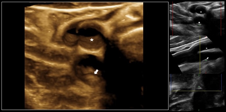
'JACC': 3D visualization of cholesterol plaques improves cardiovascular risk prediction
CNIC researchers show the value of total atherosclerosis burden for the identification of individuals at risk of cardiovascular disease
Three-dimensional ultrasound could become a central tool for identifying individuals at risk of developing cardiovascular disease. This is the conclusion of a report from the PESA study (Progression of Early Subclinical Atherosclerosis) CNIC-Santander, published in The Journal of the American College of Cardiology. The report demonstrates that total atherosclerosis burden (the amount of cholesterol accumulated in arterial walls throughout the body) is a very useful parameter in the stratification of individual cardiovascular risk, when considered together with classical risk factors (blood cholesterol, blood pressure, diabetes, smoking status, exercise, obesity).
The results of the study, directed by CNIC Director General Dr. Valentín Fuster, show that among the study participants (mean age 45 years) the total atherosclerosis burden is twice as high in men as in women (63.4 versus 25.7 cubic millimeters), is higher in the femoral arteries than in other vascular territories, and increases with age.
The CNIC scientists explored the most atherosclerosis-prone regions of the carotid and femoral arteries. In each territory, the researchers performed 3D ultrasound examinations of 6 cm arterial segments in both branches, centered on the carotid sinus and the femoral bifurcation. The study population included 3860 middle aged participants with no disease symptoms and no history of myocardial infarction, stroke, etc. All participants were Banco Santander employees based in Madrid. PESA-CNIC is a prospective cohort study that monitors patients over several years. The patients were examined with a new ultrasound system made by CNIC technology partner for the laboratory of Human Cardiovascular Imaging, Philips. The system uses a linear volumetric transducer that acquires 3D images of the arteries and atherosclerotic plaques.
Together with classical risk factors, 3D vascular ultrasound could become a central tool in the stratification of individual cardiovascular risk
“3D vascular ultrasound is a straightforward, highly reproducible, and novel imaging technique that allows the early quantification of total atherosclerosis burden in large populations,” affirmed lead author Dr. Fuster. “This new method can be used to obtain images of the atherosclerosis burden in peripheral arteries (carotid and femoral) from early to late disease stages, and can help to identify individuals at higher risk and offer them more focused and targeted treatments. However, epidemiological population studies are needed to evaluate the usefulness of this method compared with established methods in large-scale clinical practice.”
Clinical application
Although 3D ultrasound is still in the development phase, it has already shown clinical promise in several areas, including quantification of atheroscerotic plaque volume. Direct quantification of plaque volume by 3D ultrasound is more accurate than 2D techniques for estimating an individual’s total plaque burden. Cardiologist Beatriz López Melgar, a specialist in cardiovascular imaging and a researcher on the PESA study, explained some of the advantages of the new technology. “This new 3D visualization technology provides a new focus for the study of atherosclerosis and offers a plethora of new possibilities. The method allows us to evaluate the extent, severity, and characteristics of atherosclerotic plaques in 3 dimensions in just a few seconds, providing more complete information and in a simpler form than conventional 2D methods. The method also requires no patient preparation and does not involve radiation, unlike some other approaches used to study atherosclerosis and cardiovascular risk; this means that 3D ultrasound can be used in most patients without restriction and allows defined follow-up.”
"Banco Santander aims to provide the best working environment for our employees and to promote health in the general population"
Borja Ibáñez, CNIC Research Director and a cardiologist at Fundación Jiménez Díaz university hospital added that “conventional risk factors (blood cholesterol, hypertension, smoking, etc.) allow a broad prediction of the population risk of severe cardiovascular events (infarction or stroke); however, individualized predictions based on these risk factors are not very accurate. Now, with the ability to directly visualize atherosclerotic plaques and quantify their number and size in the body, we will be able to predict risk more accurately. The presence and extent of atherosclerosis gives direct information about how risk factors affect the arteries in each individual.”
The PESA study is a collaborative project between the CNIC and Banco Santander that studies more than 4000 middle aged participants with an average age of 45 years. Banco Santander Medical Services Director Dr. José María Mendiguren, an author on the study, explained the bank’s participation. “Banco Santander aims to provide the best working environment for our employees and to promote health in the general population. To achieve this, the Medical Services department is in charge of promoting health and wellbeing among our employees. The new study, published in a high-impact medical journal, is an example of the value of collaboration between the public and private sectors, represented in this case by the Pro-CNIC Foundation and with the participation of the Banco Santander Medical Services department. The study provides invaluable information, both for the individual volunteers and for the medical profession, about the early detection of this extremely important disease. Future PESA publications will provide further insights, and I hope that this study will encourage other businesses and public institutions to engage in similar projects, to the benefit of participants and society at large.”
The ambitious PESA project, launched in 2010, uses the latest diagnostic imaging technology to address unresolved questions about atherosclerosis, including when it begins and what events need to occur for it to manifest clinically. Atherosclerosis is a slow developing disease involving the accumulation of diverse materials, mostly cholesterol, in the artery walls. This accumulation can occur in any artery in the body and can lead to obstruction of the blood flow.


