News search
|
Research 17 May 2022 The 3D atlas has allowed the scientists to identify the beginning of left–right asymmetry in the heart |
|
Research 18 Apr 2022 The research will provide insights into the development of potential treatments for neurological diseases and obesity, among other human conditions |
|
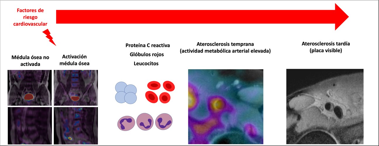
Research 21 Mar 2022 The new discovery suggests new research avenues toward the discovery of interventions to prevent or arrest atherosclerosis |
|
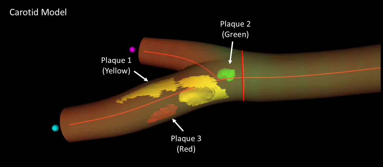
Research 16 Mar 2022 Scientists at the CNIC have led the development of a new three dimensional ultrasound method that improves the assessment of cardiovascular risk in healthy individuals |
|
Research 3 Mar 2022 Most biological processes require the import to the cell nucleus of key regulatory factors; one of the most important of these factors is the protein YAP |
|
About the CNIC 23 Feb 2022 Researchers from the CNIC and Columbia University (USA) review the role of acquired mutations and clonal hematopoiesis in cardiovascular disease |
|
About the CNIC 3 Feb 2022 |
|
Research 5 Jan 2022 Scientists at the CNIC have developed a simple model for studying the behavior of immune cells in live animals and have identified a harmful cell behavior pattern associated with cardiovascular disease |
|
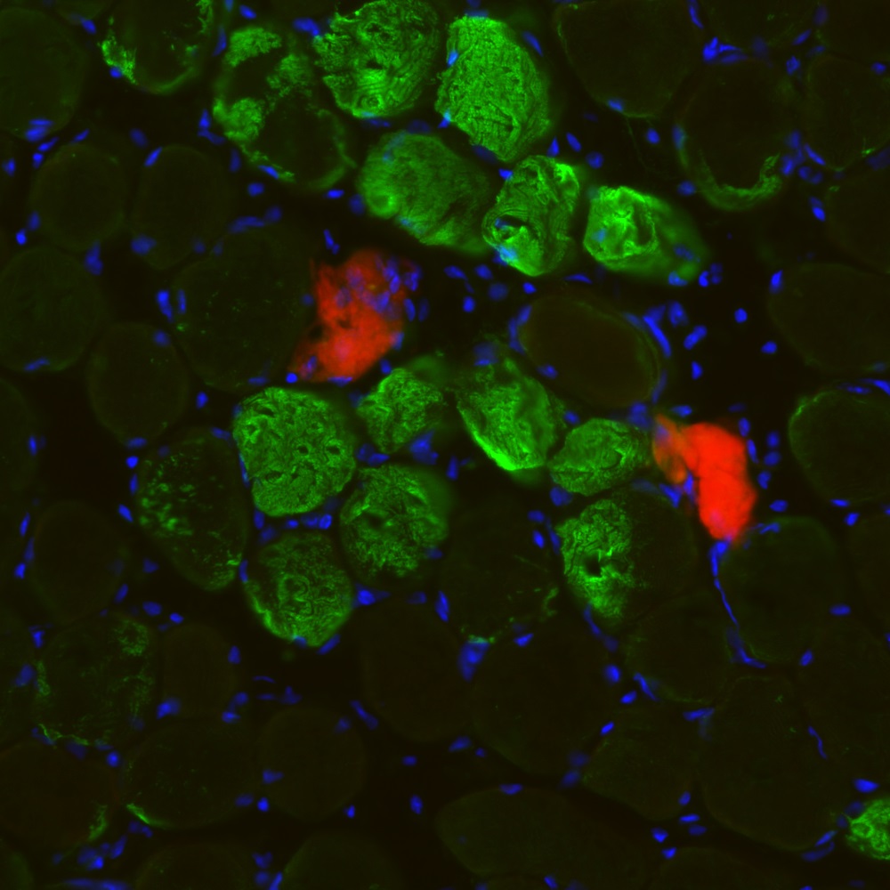
Research 18 Oct 2021 Researchers from the National Center for Cardiovascular Research and the Pompeu Fabra University describe in Science a new mechanism for muscle regeneration after physiological damage |
|
Research 3 Sep 2021 The new findings, published in Circulation Research, could spur the development of new tools for the treatment of cardiac hypertrophy |
- ‹ previous
- 5 of 24
- next ›

