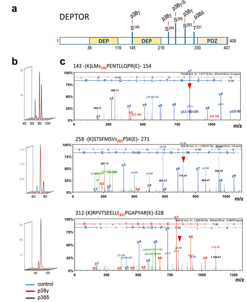
Figure 1.- Analysis of DEPTOR phosphorylation by p38γ and p38δ kinases in vivo by mass spectrometry. (a) Scheme of DEPTOR phosphorylation sites. (b) Quantitative analysis of phosphorylation. (c) MS/MS spectra of each phosphopeptide, showing its sequence and assignation of the modified site.

Figure 2.- Phosphatase SHP2 is sulfenylated in conditions of laminar shear stress (LSS). Sulfenylated SHP2 was immunoprecipitated using a specific antibody and the amount of SHP2 was quantified by mass spectrometry. Seven of the fragments of the SHP2 peptide indicated in the inset were quantified, showing that sulfenylation of SHP2 takes place when the samples were treated with H2O2 or subjected to LSS.
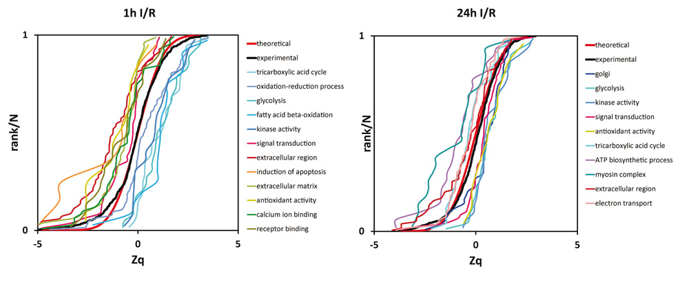
Figure 3.- Systems biology analysis of protein abundance changes in a pig infarct model. A high-throughput quantitative proteomics analysis was performed to compare heart tissue from infarcted and remote areas after 1h ischemia and 1h or 24 hreperfusion. The analysis revealed a clearly coordinated behavior of proteins belonging to the indicated functional categories.


