Cells
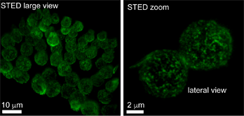
3D-gSTED rendering of 4µm Z-stacks of human leucocytes immunostained for the HS1 protein.
Sampietro M, Zamai M, Díaz Torres A, Labrador Cantarero V, Barbaglio F, Scarfó L, Scielzo C and Caiolfa V. Front. Cell Dev. Biol. 2021.
Tracking of CD3β-mCherry vesicles in Jurkat cells by Total Internal Reflection Microscopy (TIRFM).
Blas-Raus N., et al., Nat. Communication, 2016.
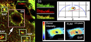
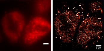
Interaction of proteins (in magenta) of Endothelial Adhesive Platforms (EAPs) at the plasma membrane detected by 2-photon phasor FLIM-FRET analysis.
Barreiro O., et al., J Cell Biol. 2008.
Tissues
Light sheet fluorescence image (Bruker SPIM LCS) of a clarified mouse heart. Microvasculature (white), macrovasculature (red), lineage-traced cell nuclei (green).
Luis Diago (CNIC) 2024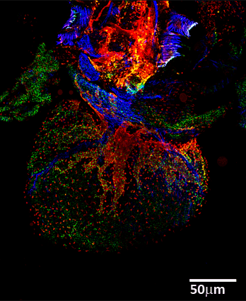
Tiled High Resolution Confocal Image of a cleared mouse embryonic heart. SMAcy3 (blue), Lyvel 633 (red) and Prox1 cy5.5 (green).
Lioux G., et al., Dev Cell 2020.
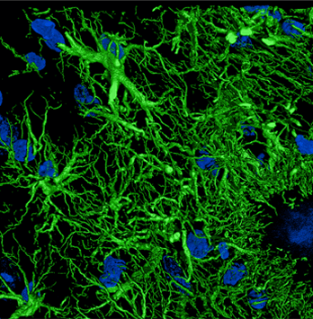
Hypertrophic and reactive astrocytes (GFAP-green) imaged by Confocal microscopy and processed by Huygens deconvolution.
Marta Cortes Canteli (CNIC) 2019.
Organisms
2-photon microscopy time-lapse video of a Mesp1cre/+;Rosa26Rtdtomato+/- embryo -reporting anterior mesoderm-, showing the transition from cardiac crescent stage to heart tube stage.
lvanovitch K., et al. eLife 2017.
Oblique illumination and dSTORM super-resolution images of mouse pre-implantation embryo nuclei, H3K4me3 histone Alexa 647 Ab..
Marta Portela Martinez and Maria José Andreu (CNIC) 2019.


